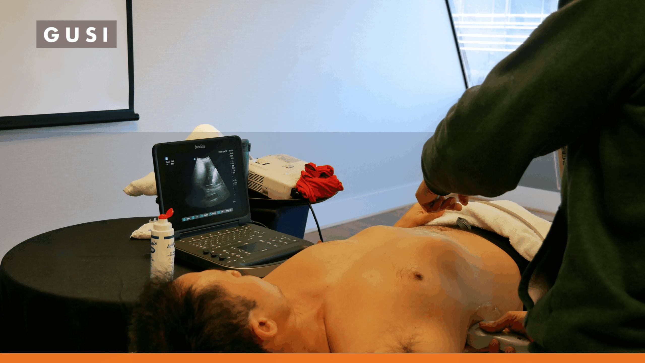


Shortness of breath, cough, hypoxemia, chest discomfort, and abnormal lung sounds can come from very different problems. Pneumonia, pulmonary edema, pleural effusion, pneumothorax, asthma, COPD, pulmonary embolism, and nonpulmonary disease can overlap clinically, especially early in the evaluation.
Lung point-of-care ultrasound, or lung POCUS, gives trained clinicians a rapid bedside way to look for patterns that narrow that differential. It is particularly useful because many lung diseases change the relationship between air, fluid, pleura, and peripheral lung tissue in ways ultrasound can detect.
The Direct Answer
Lung POCUS supports respiratory diagnosis by identifying focused ultrasound patterns associated with pneumonia, pulmonary edema, pleural effusion, pneumothorax, and other causes of acute dyspnea. It does not identify a respiratory virus, confirm every pneumonia, rule out pulmonary embolism, or replace CT and other imaging when a broader evaluation is needed.
The most useful question is not, ‘What disease does this one artifact mean?’ It is, ‘What pattern is present, how is it distributed, and does it fit the patient’s clinical picture?’
What Is Lung POCUS?
Normal aerated lung reflects and scatters ultrasound at the pleural interface, so clinicians often interpret artifacts rather than seeing deep lung tissue directly. When air is replaced or interrupted by fluid, consolidation, or other pathology near the pleural surface, the ultrasound pattern changes.
Lung POCUS uses those changes to answer focused questions at the bedside. The exam can be repeated during the same encounter, performed without ionizing radiation, and integrated immediately with vital signs, physical examination, cardiac POCUS, laboratory results, and treatment response.
The technique is powerful but deliberately limited. Central pathology that does not reach the pleura may be missed, and the same ultrasound sign can have several causes.
How to Perform a Lung POCUS Exam
Choose the Probe for the Question
A curvilinear or phased-array transducer provides depth and a broad field for general lung assessment, consolidation, and pleural effusion. A high-frequency linear transducer provides excellent detail of the pleural line and is especially useful when evaluating lung sliding or a small pneumothorax.
Different machines and presets change the appearance of artifacts, so clinicians should use a consistent setup and understand how their device handles harmonics, compounding, depth, focal zone, and gain.
Scan Multiple Lung Zones
A limited anterior scan can answer a very specific question, but a diagnostic respiratory examination usually benefits from systematic bilateral scanning. Common protocols divide each hemithorax into anterior, lateral, and posterior zones, with multiple interspaces examined in each region.
Posterior and dependent zones matter in pneumonia and pleural effusion. If the patient’s condition allows, repositioning can improve access to those areas.
Key Lung Ultrasound Findings
| Finding | What It Looks Like | What It Can Mean |
| A-lines | Horizontal repeating artifacts parallel to the pleural line | Normal aerated lung, obstructive physiology such as asthma/COPD, or pneumothorax when combined with loss of pleural apposition signs. |
| B-lines | Vertical hyperechoic artifacts arising from the pleural line and extending through the image | Increased peripheral lung density / interstitial-alveolar involvement. Diffuse bilateral patterns may support edema; focal patterns can occur with pneumonia, contusion, infarction, and other disease. |
| Lung sliding | Shimmering movement at the pleural interface with respiration | Presence supports apposition of visceral and parietal pleura at that location and argues against pneumothorax there. |
| Lung pulse | Pleural movement synchronized with cardiac activity when ventilation-related sliding is absent | Shows pleural apposition and argues against pneumothorax at that location. |
| Lung point | Transition between pneumothorax pattern and normally apposed lung during respiration | Highly specific for pneumothorax when present, though it may not be found in a very large pneumothorax. |
| Consolidation | Tissue-like or hypoechoic peripheral lung replacing the normal artifact pattern | Pneumonia, atelectasis, contusion, infarction, or other peripheral lung pathology. |
| Dynamic air bronchograms | Bright branching air echoes moving with respiration inside consolidation | Support pneumonia over resorptive atelectasis in the right clinical context, but are not present in every pneumonia. |
| Pleural effusion | Anechoic or complex fluid between pleural surfaces above the diaphragm | Transudate, exudate, blood, pus, or other pleural fluid. Internal echoes and septations may suggest complexity but do not determine etiology alone. |
| Pleural-line irregularity | Irregular, thickened, or fragmented pleural appearance | Can occur with inflammatory, fibrotic, infectious, or other peripheral lung processes; inter-reader agreement can be limited. |
POCUS for Pneumonia
Pneumonia is one of the strongest evidence-supported uses of lung ultrasound. Peripheral consolidation can appear tissue-like or ‘hepatized,’ often with an irregular shred-like deep border, focal B-lines, and air bronchograms.
In 2025, the American Thoracic Society issued a conditional recommendation that lung ultrasound is an acceptable diagnostic alternative to chest radiography for adults with suspected community-acquired pneumonia in centers where appropriate clinical expertise exists. The guideline also identifies situations that weaken that recommendation, including concern for alternative diagnoses such as pulmonary embolism or malignancy and technical barriers to high-quality scanning. [1]
A 2024 meta-analysis of 29 studies and 6,702 adults reported pooled sensitivity of 92% and specificity of 94% for lung ultrasound diagnosis of pneumonia. [2] An updated 2025 comparison found better pooled diagnostic performance for LUS than chest radiography in the included CAP studies, though heterogeneity was substantial. [3]
Pneumonia Guardrail
Lung ultrasound is best at detecting pneumonia that reaches the pleural surface. A central pneumonia surrounded by normally aerated lung can be invisible to ultrasound. A normal scan therefore does not eliminate every pneumonia in every patient.
POCUS for Pulmonary Edema and Acute Heart Failure
Diffuse bilateral B-lines are a key lung ultrasound pattern in acute pulmonary congestion. When they are symmetric, present in multiple zones, and fit the clinical picture, they can strongly support cardiogenic pulmonary edema.
A 2025 meta-analysis of 38 studies including 6,783 patients reported pooled sensitivity of 92% and specificity of 90% for lung ultrasound in heart failure, while also finding high heterogeneity across studies. [4]
B-lines should not be interpreted as ‘heart failure lines.’ Interstitial pneumonia, ARDS, pulmonary fibrosis, contusion, and other processes can produce vertical artifacts. The distribution, pleural appearance, symptoms, cardiac findings, and treatment response all matter.
For undifferentiated dyspnea, pairing lung ultrasound with focused cardiac POCUS can be more informative than interpreting the lungs alone.
POCUS for Pleural Effusion
Pleural effusion is usually straightforward to identify with lung ultrasound because fluid creates an acoustic window where aerated lung normally blocks transmission. The scan can show the diaphragm, adjacent lung, fluid characteristics, and sometimes internal septations or echogenic material.
ACEP’s Lung Sonoguide cites sensitivity of approximately 88%-94% and specificity of 96%-100% across older studies, with ultrasound outperforming supine chest radiography for detecting pleural fluid. [5]
Ultrasound can also guide thoracentesis, but procedural planning requires attention to diaphragm position, lung movement, fluid depth, patient position, and local procedural protocols.
POCUS for Pneumothorax
Pneumothorax changes the pleural interface by separating the visceral pleura from the chest wall. Common ultrasound findings include absent lung sliding, absent B-lines, and a stratosphere/barcode pattern on M-mode. A lung point, when present, is strongly supportive of pneumothorax.
The important safety point is that absent sliding is not specific. Apnea, mainstem intubation, severe atelectasis, pleurodesis, fibrosis, phrenic nerve dysfunction, and other conditions can also reduce or eliminate visible sliding. [5]
Conversely, visible B-lines, a lung pulse, or clear lung sliding at a scanned location indicate visceral and parietal pleural apposition and therefore argue against pneumothorax at that point.
Clinical urgency matters. In an unstable patient with suspected tension pneumothorax, ultrasound should not delay life-saving treatment.
What About COPD and Asthma?
Lung POCUS is usually more helpful for finding an alternative cause of dyspnea than for directly diagnosing an asthma or COPD exacerbation.
A patient with obstructive disease may show an A-line predominant pattern with preserved lung sliding and no focal consolidation or diffuse B-line syndrome. That pattern can fit asthma or COPD, but it is not specific. A similar A-line pattern can occur in normal lung.
The value is often what the scan does not show: no pulmonary edema pattern, no pleural effusion, no focal peripheral pneumonia, and no pneumothorax pattern. The diagnosis of asthma or COPD exacerbation remains clinical and may require spirometry or other testing outside the acute setting.
Can Lung POCUS Diagnose Pulmonary Embolism?
No. Lung POCUS cannot reliably exclude pulmonary embolism.
Some emboli cause peripheral pulmonary infarcts that reach the pleura and can appear as small wedge-shaped consolidations, sometimes with pleural fluid. Focused cardiac POCUS may show right-heart strain in larger pulmonary emboli, and compression ultrasound may identify a deep-vein thrombosis.
Those findings can raise or lower suspicion in the right setting, but they do not replace validated PE pathways, D-dimer testing, CT pulmonary angiography, ventilation-perfusion imaging, or specialist assessment when indicated.
POCUS During Cold, Flu, RSV, and COVID Season
Seasonal respiratory viruses are an important reason patients present with cough and dyspnea, but lung ultrasound does not identify which virus is causing the illness.
An uncomplicated upper respiratory infection or mild influenza-like illness usually does not need lung POCUS simply because the patient has a cough. The exam becomes more useful when the clinical question shifts to lower-respiratory or cardiopulmonary complications:
- Is there focal consolidation concerning for pneumonia?
- Is there a pleural effusion?
- Are there diffuse B-lines suggesting pulmonary congestion or a diffuse interstitial process?
- Is pneumothorax a concern?
- Is the lung scan unrevealing enough that the clinician should broaden the evaluation to the heart or another system?
COVID-era literature helped expand lung-ultrasound experience, but findings such as B-lines, subpleural consolidations, and pleural irregularity are not specific to COVID-19. Viral testing remains the appropriate way to identify a respiratory virus when knowing the pathogen matters.
The seasonal angle therefore belongs inside the respiratory guide, not around it.
Use Cardiopulmonary POCUS for Undifferentiated Dyspnea
Respiratory symptoms are not always caused by the lungs. Pericardial effusion, left- or right-heart dysfunction, volume overload, and other cardiovascular problems can present with cough, chest discomfort, tachypnea, or exercise intolerance.
When the lung scan does not explain the severity of the symptoms, a focused cardiac examination can be a natural next step for a clinician trained in cardiopulmonary POCUS.
| Lung Pattern | Possible Next Question |
|---|---|
| Diffuse bilateral B-lines | Does focused cardiac POCUS support cardiogenic congestion, or is another diffuse lung process more likely? |
| Focal consolidation | Does the clinical picture support pneumonia, or are atelectasis, infarction, contusion, or another cause plausible? |
| Pleural effusion | Is the effusion part of heart failure, infection, malignancy, trauma, or another process? |
| A-line pattern with ongoing severe dyspnea | Is there obstructive disease, pulmonary embolism, cardiac disease, metabolic illness, or another non-lung explanation? |
| Lung findings mild/unrevealing | Does focused cardiac POCUS reveal pericardial effusion, ventricular dysfunction, or right-heart strain? |
When Lung POCUS Is Not Enough
The correct endpoint of a POCUS examination is sometimes ‘I need more information.’ Chest radiography, CT, echocardiography, laboratory testing, microbiology, or specialist imaging may still be needed when:
- The ultrasound exam is technically limited or important lung regions cannot be visualized.
- Symptoms are severe or worsening despite a low-risk or nondiagnostic lung scan.
- A central pneumonia, mass, pulmonary embolism, aortic disease, or other deep thoracic process remains possible.
- Detailed anatomic characterization or disease staging is required.
- The clinical picture and ultrasound findings do not match.
- A finding needs formal measurement, surveillance, or specialist interpretation.
- The patient is unstable and definitive imaging or treatment must not be delayed.
Common Pitfalls and Limitations
- Calling every B-line pattern pulmonary edema. Distribution and clinical context are essential.
- Calling absent lung sliding a pneumothorax. Look for B-lines, lung pulse, lung point, and alternative causes.
- Scanning only the anterior chest when dependent pneumonia or effusion is plausible.
- Using one positive interspace to label a diffuse process.
- Assuming no consolidation means no pneumonia. Central lesions may not contact the pleura.
- Treating static vs dynamic air bronchograms as infallible. They modify probability rather than create certainty.
- Overinterpreting pleural-line irregularity, which has inter-reader variability.
- Forgetting that probe choice, machine settings, body habitus, dressings, wounds, subcutaneous emphysema, and positioning can change image quality.
- Failing to archive or document representative images when local policy, QA, credentialing, or billing requires it.
Training and Competency
Lung ultrasound is often one of the first POCUS applications clinicians learn because normal patterns and major artifacts are visually memorable. Clinical integration is harder.
Competency includes recognizing when the exam is appropriate, scanning enough lung zones, acquiring reproducible images, distinguishing artifacts from pathology, integrating the distribution of findings, knowing important mimics, documenting limitations, and deciding when to escalate.
Current international consensus also emphasizes standardized terminology and examination protocols to improve reproducibility. [6]
A short course can build the foundation, but continued scanning and image review are what reduce pattern-recognition errors over time.
Lessons From Two Original GUSI Cases
The December 2023 article included two cases from GUSI instructor Sebastian Schoneich, MD. They still teach useful lessons when framed carefully.
Case 1: Respiratory Symptoms With an Unrevealing Lung Scan
A 63-year-old man presented with dry cough and intermittent chest discomfort. The lung examination showed no major pulmonary edema, effusion, or large consolidation. His discomfort worsened when he lay flat, prompting a focused cardiac exam.
The parasternal long-axis and subxiphoid views showed a significant pericardial effusion with concern for early tamponade physiology, and the patient underwent drainage.
The enduring lesson is not that every patient with a cough needs an echocardiogram. It is that a respiratory complaint with a lung scan that does not explain the clinical picture should prompt the clinician to reconsider the differential, including cardiac causes.
Case 2: More Than One Diagnosis Can Be Present
A 68-year-old man with known heart failure presented with cough, worsening exertional dyspnea, edema, fever, and weight gain. Lung ultrasound showed diffuse B-lines consistent with pulmonary congestion, but also a focal left lower-lung consolidation with a shred-like border and a small pleural effusion.
Those additional findings increased concern for a concurrent focal pulmonary process rather than isolated heart-failure exacerbation.
The lesson is that POCUS can reveal overlapping patterns. Diffuse B-lines do not prevent a patient from also having pneumonia, and focal consolidation does not eliminate coexisting congestion. Management should follow the full clinical assessment and, when needed, additional imaging and testing.
Updated Expert Bio: Sebastian Schoneich, MD
Sebastian Schoneich, MD, is a GUSI instructor and family physician. GUSI currently lists him as an Assistant Clinical Professor in Family Medicine & POCUS at the University of Texas at Austin Dell Medical School and a staff physician at a federally qualified health center in Austin, Texas. He completed an Advanced Primary Care POCUS fellowship at the University of Michigan and has extensive experience using POCUS in resource-limited primary care settings. Dr. Schoneich’s professional interests encompass global health and health equity, an exploration of the broader structural sources of diseases, and the education and application of POCUS to diminish barriers to care in resource-limited primary care settings.
Beyond his professional endeavors, Sebastian enjoys cycling, daydreaming about the seaside, and spending quality time with family and friends at a beer garden