
Point-of-care ultrasound (POCUS) and traditional diagnostic sonography can look very different in practice, but they are built on the same ultrasound technology. Both use sound waves and a transducer to create real-time images. What changes is why the examination is being performed, who performs and interprets it, how much anatomy is evaluated, how the study is documented, and how the result fits into the patient’s care.
The Direct Answer
POCUS is a focused ultrasound examination performed at the point of care to answer a specific clinical question or guide a procedure. Traditional diagnostic sonography is generally a more comprehensive, protocol-driven examination performed within a formal imaging workflow. Neither is inherently ‘better.’ The right approach depends on the clinical question, operator training, required level of detail, and whether a focused answer is sufficient.
For clarity, this guide uses the phrase “traditional sonography” because that is how many people search for the comparison. Clinically, “comprehensive diagnostic ultrasound” or “diagnostic sonography” is usually the more precise description.
POCUS vs. Traditional Sonography at a Glance
| Comparison | POCUS | Comprehensive Diagnostic Sonography |
|---|---|---|
| Primary purpose | Answer a focused clinical question or guide a procedure | Perform a comprehensive diagnostic examination using an established protocol |
| Typical operator | Treating clinician or other appropriately trained licensed medical provider | Diagnostic medical sonographer, physician, or other qualified imaging professional depending on the exam and setting |
| Interpretation | Often interpreted immediately by the clinician performing the exam | Images/findings generally enter a formal interpretation and reporting workflow |
| Scope | Limited and question-specific | Broader, protocol-driven, and designed for diagnostic completeness |
| Timing | Integrated into the current encounter | Performed as an imaging examination, sometimes immediately and sometimes scheduled |
| Location | Where the patient is being cared for: bedside, clinic, field, procedure room, etc. | Imaging department, vascular lab, clinic, hospital room, mobile service, or other equipped setting |
| Equipment | Handheld, portable, cart-based, or full-size ultrasound system | Portable, cart-based, or full-size ultrasound system with application-specific capabilities |
| Documentation | Focused findings, images/clips, limitations, interpretation, and clinical action according to local policy | Protocol images, measurements, technical documentation, and formal report according to specialty/facility standards |
| Quality framework | Training, scope, competency, image review, QA, and escalation | Training, credentialing, protocol compliance, interpreting-provider standards, QA, and accreditation where applicable |
| Best fit | Time-sensitive bedside decisions and focused questions | Comprehensive anatomy, formal measurements, complete diagnostic evaluation, or specialist-level detail |
First, They Use the Same Ultrasound Technology
The easiest misconception to clear up is that POCUS is not a separate type of ultrasound machine. A handheld device can be used for POCUS, but a full-size system can also be used at the point of care. Likewise, a portable machine can be used to perform a formal diagnostic study.
What makes an examination POCUS is the workflow: ultrasound is used by the treating team at the point of care to answer a defined question or guide an intervention, and the result is integrated immediately into clinical decision-making. The American Institute of Ultrasound in Medicine (AIUM) describes POCUS as bringing ultrasound to the bedside for real-time visualization, assessment, and treatment decisions. [1]
So the distinction is less about the machine and more about the question being asked.
What Is Traditional Diagnostic Sonography?
Diagnostic medical sonography is a professional imaging discipline with its own body of knowledge, educational standards, credentials, specialty areas, and quality expectations. The Society of Diagnostic Medical Sonography defines the sonographer as a professional who performs diagnostic sonographic examinations and provides the resulting images, findings, or results to an interpreting physician or other legally authorized provider. [2]
A comprehensive diagnostic examination is typically performed according to a defined protocol. That may include multiple views, measurements, Doppler data, comparison with prior imaging, and documentation required by the specialty or facility. The goal is not simply to answer one narrow bedside question; it is to create a diagnostic examination that is complete enough for formal interpretation.
The exact workflow varies. Some examinations are performed in imaging departments, while others happen in clinics, vascular labs, patient rooms, mobile services, operating rooms, or physician offices. Some results are interpreted immediately, while others move through a scheduled reporting process. Those differences are why ‘traditional sonography takes days’ is not a reliable distinction.
What Is POCUS?
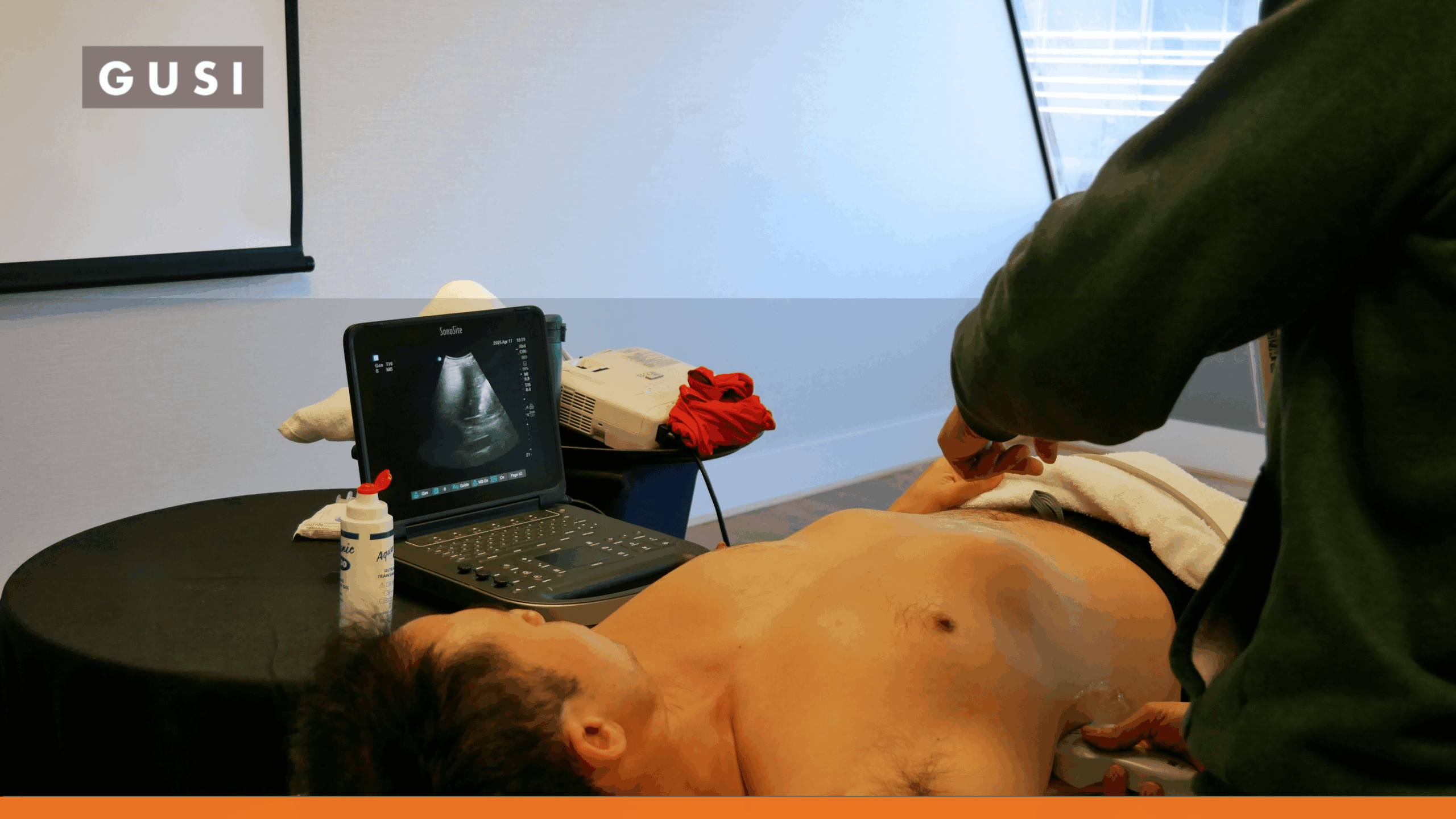
POCUS is ultrasound used as part of the clinical encounter. The operator starts with a focused question such as: Is there free fluid? Is the bladder distended? Is there a pericardial effusion? Is there lung sliding? Is the procedure target where I expect it to be?
The clinician acquires the images, interprets them in the context of the patient, and uses the answer to inform the next action. The exam may take minutes, but ‘quick’ does not mean casual. A focused study still needs appropriate technique, documentation, recognition of limitations, and a plan for what happens when the exam is negative, technically limited, or discordant with the clinical picture.
The Biggest Differences: Purpose, Scope, Workflow, and Responsibility
Focused Question vs. Comprehensive Examination
POCUS succeeds when it answers the question it was designed to answer. A comprehensive diagnostic study succeeds when it evaluates the required anatomy and measurements across the full protocol. That is a different standard of completeness.
A clinician looking for hydronephrosis in a patient with flank pain does not necessarily need to reproduce an entire renal ultrasound protocol to make the next bedside decision. But if the question is renal anatomy, mass characterization, vascular detail, or a comprehensive diagnostic assessment, a formal study may be necessary.
Clinician-Integrated Interpretation vs. Formal Imaging Workflow
One of POCUS’s defining strengths is immediacy. The person holding the probe often already knows the patient’s history, examination findings, vital signs, and working differential. The ultrasound image becomes one more piece of the bedside examination.
Comprehensive sonography brings a different strength: systematic acquisition designed to support formal interpretation. Sonographers contribute deep technical expertise in anatomy, optimization, protocols, measurements, artifacts, and pathology recognition. The interpreting provider can review a standardized body of images rather than a single focused answer.
Protocol, Documentation, and Reporting
Both workflows require documentation. AIUM’s current training guidelines for physicians and advanced clinical providers include methods of documenting and reporting ultrasound studies, and current AIUM accreditation standards include requirements for quality assurance, reports, equipment, and record keeping. [3][4][5]
The documentation may look different because the exams serve different purposes. A focused POCUS note may record the indication, views obtained, key findings, limitations, interpretation, and resulting clinical action. A comprehensive sonographic study is generally documented through a formal imaging protocol and report.
Equipment and Location Do Not Define POCUS
The original newsletter contrasted handheld POCUS with larger traditional machines. That was useful shorthand, but modern practice is more flexible. POCUS may be performed with a handheld probe, a compact laptop-style unit, a cart-based system, or a full-featured ultrasound platform. Comprehensive diagnostic sonography can also be performed on portable equipment.
The better distinction is purpose and workflow, not device size.
A Sonographer-Turned-PA Perspective
Agnes Lovell brings a rare perspective to this comparison. Before becoming a PA-C, she spent seven years in vascular sonography, including work as a clinical instructor and lead. She then entered PA training and began using ultrasound in environments such as emergency care, surgery, procedures, and internal medicine.
For someone trained to pursue technically complete, protocol-driven images, the shift to focused POCUS can feel almost counterintuitive. Agnes described having to let go of the instinct to make every exam look like a comprehensive diagnostic study and instead ask whether the images were adequate to answer the clinical question.
Agnes Lovell, PA-C, RVT
“POCUS is much more practical and answers specific clinical questions.”
What a Sonographer May Have to ‘Unlearn’
The transition is not about lowering standards. It is about changing the definition of success. In diagnostic sonography, the operator may work systematically through a complete protocol even when some images are technically difficult. In POCUS, the clinician may stop once a validated focused question has been answered or recognize quickly that the study is nondiagnostic and escalate.
For a sonographer, that can initially feel incomplete. For a clinician, it can be exactly what the encounter requires.
What Sonography Expertise Brings to POCUS
Experienced sonographers bring skills that are enormously valuable to POCUS education: probe control, anatomy, image optimization, Doppler, artifact recognition, standardized acquisition, ergonomics, and an instinct for when an image is not good enough.
That expertise is especially valuable in training programs and quality assurance. POCUS should not grow by dismissing the craft of sonography; it should borrow from it.
What Clinician-Performed POCUS Adds
The clinician brings the immediate clinical context and responsibility for the next decision. That creates a different feedback loop: scan, interpret, treat or triage, then reassess. POCUS can therefore become an extension of the physical examination rather than a separate diagnostic event.
The strongest systems combine both strengths: sonographic image quality and discipline with clinician-centered integration and speed.
Is POCUS Less Accurate Than Traditional Sonography?
There is no single accuracy number for ‘POCUS versus sonography.’ Accuracy depends on the application, patient, operator, protocol, equipment, reference standard, and what the exam is expected to detect.
A focused POCUS exam can be highly accurate for a narrow, well-validated question in trained hands. But a focused exam is intentionally less comprehensive. It may never look for pathology outside its scope.
That means a negative POCUS result is only meaningful when the application has sufficient sensitivity for the clinical question and the study was technically adequate. A comprehensive diagnostic study is usually better when the clinician needs a broader evaluation, formal measurements, detailed anatomy, or pathology characterization.
Better Question
Instead of asking “Which is more accurate?”, ask: “What question must this examination answer, how complete does the evaluation need to be, and what should happen if the focused study is negative or limited?”
When POCUS May Be Enough
Depending on the application, training, and local scope, POCUS may provide enough information to make the next clinical decision when:
- The question is focused and validated for bedside ultrasound.
- The operator is trained and competent in the application.
- Adequate views are obtained and the limitations are understood.
- The result fits the clinical picture.
- The examination changes immediate treatment, triage, or procedure planning.
- There is a clear follow-up or escalation plan if symptoms persist or the patient’s condition changes.
When Comprehensive Diagnostic Ultrasound Is Still Needed
A formal diagnostic study is usually the better next step when:
- A comprehensive anatomic survey or complete specialty protocol is required.
- Formal measurements, Doppler characterization, serial surveillance, or detailed pathology characterization are needed.
- The POCUS exam is technically limited or nondiagnostic.
- The focused result does not match the patient’s symptoms or examination.
- An unexpected or incidental abnormality needs characterization.
- The diagnosis has important management consequences that require formal confirmation.
- Local policy, scope of practice, accreditation, credentialing, reimbursement, or specialist workflow requires a formal study.
Training: POCUS Does Not Mean Minimal Training
A smaller exam scope does not mean the operator needs only a smaller commitment to quality. Current AIUM guidance for physicians and advanced clinical providers emphasizes indications, limitations, ultrasound physics, anatomy and pathophysiology, image analysis, documentation, reporting, procedural experience, and maintenance of competence. [3][4]
That is important for PAs and other advanced practice providers. AIUM’s current training guidance specifically includes PAs, NPs, midwives, physical therapists, and other licensed provider groups within defined pathways, subject to active licensure, relevant scope, supervised training, experience, and maintenance requirements. [4]
For health systems, competency also needs infrastructure: image archiving, review, documentation standards, quality assurance, equipment maintenance, and escalation. The technology is portable. Responsibility is not.
How POCUS and Sonography Work Better Together
POCUS works best when it is not framed as a replacement for sonographers or radiology. It answers a different layer of the clinical problem.
| POCUS Can… | Comprehensive Sonography Can… | Together They Can… |
|---|---|---|
| Answer a focused bedside question immediately | Provide a complete diagnostic examination | Use POCUS to triage and a formal study when greater detail is needed |
| Guide treatment or a procedure in real time | Provide standardized measurements and documentation | Reduce unnecessary delay without sacrificing diagnostic completeness |
| Identify when more imaging is needed | Characterize the finding comprehensively | Create a cleaner, better-targeted referral pathway |
| Support serial bedside reassessment | Provide formal surveillance studies | Match the imaging workflow to the clinical question |
| Bring ultrasound into more clinical settings | Maintain deep sonographic expertise and protocol discipline | Strengthen training, QA, and patient safety |
For Agnes, the sonographer-to-PA transition ultimately shows why this is not an either-or debate. Diagnostic sonography taught her how to create and judge high-quality ultrasound images. POCUS gave those skills a new role inside the clinical encounter.
The best POCUS clinicians know when a focused answer is enough. The best ultrasound systems also make it easy to call on comprehensive sonography when it is not.
Build the Skills Behind the Image
POCUS is useful because it is focused, not because it is effortless. GUSI’s POCUS Essentials training helps clinicians build foundational acquisition, interpretation, and clinical-integration skills. Expert Scan Reviews and ScanHub can support ongoing feedback, image review, and competency development.