
Fasting instructions are designed to reduce pulmonary aspiration risk before anesthesia and deep sedation. The problem is that time since the last meal does not always tell clinicians what is actually in the stomach. Gastric emptying can be delayed by illness, medications, pregnancy, diabetes, pain, opioids, and other factors, and sometimes the fasting history is simply uncertain.
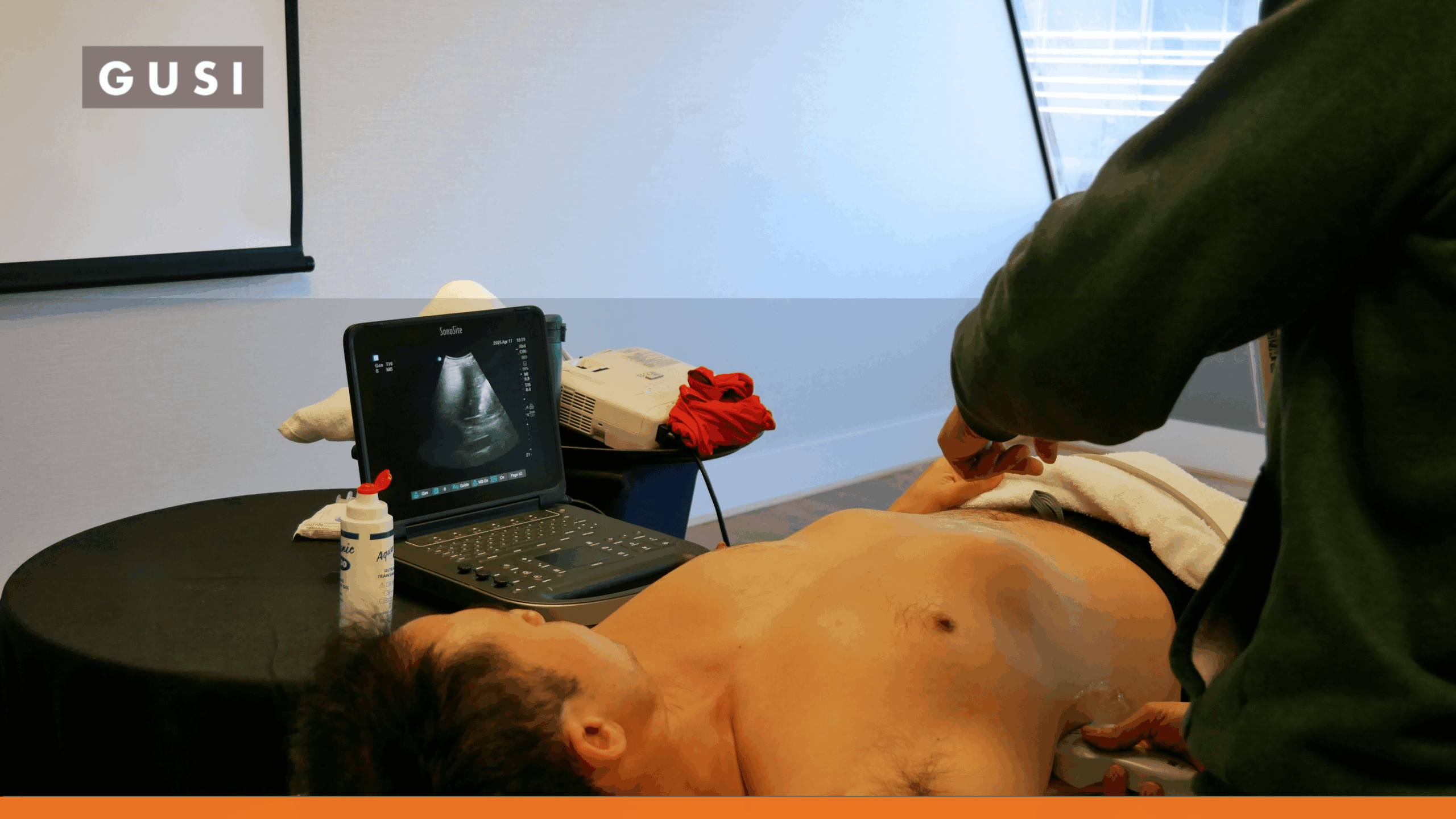
Gastric point-of-care ultrasound, or gastric POCUS, gives trained clinicians a bedside way to look directly at the gastric antrum, characterize its contents, and, when clear fluid is present, estimate gastric fluid volume.
The Direct Answer
Gastric POCUS is a focused bedside ultrasound examination used to assess whether the stomach appears empty or contains clear fluid, thick liquid, or solid material. It is most useful when aspiration risk is uncertain, such as unclear fasting status or suspected delayed gastric emptying. The findings can support decisions about procedural timing, airway management, and aspiration precautions, but they must be integrated with clinical context and local policy.
What Is Gastric POCUS?
Gastric POCUS focuses on the gastric antrum because it is the part of the stomach that can be visualized most consistently and correlates with gastric content and clear-fluid volume. The examination is usually performed with a low-frequency curvilinear transducer in an epigastric parasagittal plane. [1][2]
The exam has two parts. The first part is a qualitative assessment: does the stomach appear empty, contain clear fluid, or contain thick/solid content? Second, if the stomach contains clear fluid and the patient fits an appropriate quantitative model, the clinician may measure the antral cross-sectional area and estimate gastric fluid volume.
That sequence matters. Volume formulas should not be used to “quantify” solid food.
When Is Gastric POCUS Clinically Useful?
Gastric POCUS is not intended to replace routine fasting guidance for every healthy elective patient. Its highest value is when the clinician is uncertain whether the stomach is likely to be empty or when the consequences of an incorrect assumption are important.
| Clinical Situation | Why Gastric POCUS May Help | Important Caveat |
|---|---|---|
| Uncertain fasting history | Provides objective information when the timing or amount of intake is unclear. | A technically limited exam should not be treated as reassuring. |
| Diabetes / gastroparesis risk | May identify retained gastric contents despite standard fasting. | Interpret within the patient’s symptoms, disease severity, and procedure urgency. |
| GLP-1 receptor agonist use | Can help assess the stomach in selected higher-risk patients when concern for delayed emptying remains. | Current multi-society guidance does not recommend routine drug cessation for every patient. |
| Urgent or emergency procedure | May help refine aspiration-risk planning when time-based fasting cannot be achieved. | Urgency may still require proceeding with full-stomach precautions regardless of ultrasound. |
| Pregnancy / labor | May provide individualized information where gastric emptying and fasting status are uncertain. | Pregnancy-specific positioning, formulas, and recommendations differ from nonpregnant adults. |
| Enteral feeding or complex medical conditions | May provide bedside evidence when standard fasting assumptions are not applicable. | Evidence and recommendations vary by population. |
American Society of Regional Anesthesia (ASRA) Pain Medicine’s 2025 expert recommendations support gastric POCUS in selected medically complex groups, with stronger support for active labor, urgent cesarean delivery, and diabetes, and conditional support in settings such as obesity, emergency care, enteral feeding, and GLP-1 receptor agonist use. The same review emphasizes that gastric POCUS complements clinical judgment rather than replacing it. [3]
How to Perform a Gastric POCUS Exam
1. Choose the Probe and Depth
For most adults, use a low-frequency curvilinear transducer, typically around 2-5 MHz, with an abdominal preset or similar general abdominal setting. Set the depth so the aorta or vertebral body can be seen posteriorly. Smaller children may require a higher-frequency transducer. [2]
2. Position the Patient
A complete adult gastric POCUS assessment generally includes both the supine and right lateral decubitus positions. The right lateral decubitus position is especially important because gravity moves gastric contents toward the antrum, making small volumes easier to detect and allowing quantitative clear-fluid assessment. [2][4]
If the patient cannot be positioned safely, the exam may be limited and the interpretation should say so.
3. Find the Gastric Antrum
Place the transducer in the epigastrium in a parasagittal orientation with the marker directed cephalad. Identify the left lobe of the liver anteriorly and the pancreas and major vessels posteriorly. The gastric antrum lies between the liver and the pancreas, near the aorta or superior mesenteric artery in the standard view. [2]
The antrum changes shape with peristalsis, so measurements should be taken between contractions and from serosa to serosa.
4. Scan Supine and Right Lateral Decubitus
Start with a qualitative scan in the supine position, then repeat in right lateral decubitus. A stomach with obvious solid or thick contents may already appear full while supine. However, an apparently empty supine antrum does not exclude a meaningful volume of clear fluid. [2][4]
How to Interpret Gastric Contents
| Finding | Typical Sonographic Appearance | Interpretation |
|---|---|---|
| Empty / near-empty antrum | Small or collapsed “bull’s-eye” appearance with opposing walls and visible wall layers | Compatible with an empty stomach when confirmed in both standard positions. |
| Clear fluid | Anechoic or hypoechoic homogeneous content; recent ingestion may create small echogenic air bubbles | Requires qualitative grading; quantitative volume estimation may be appropriate in validated populations. |
| Thick liquid | More homogeneous echogenic content than clear fluid | Treated as inconsistent with an empty stomach in perioperative risk assessment. |
| Solid content | Heterogeneous or “frosted-glass” echogenic pattern; posterior structures may be obscured by air/food | Considered a full-stomach finding in common perioperative gastric POCUS frameworks. Do not apply the clear-fluid volume formula. |
Perlas Grading: Grade 0, 1, and 2
When the stomach contains no solids or thick liquid, a simple qualitative grading system can help describe clear-fluid distribution:
| Grade | Supine | Right Lateral Decubitus | Typical Meaning |
|---|---|---|---|
| Grade 0 | No clear fluid | No clear fluid | Empty / negligible contents. |
| Grade 1 | No clear fluid | Clear fluid visible | Common in appropriately fasted adults and usually reflects a low volume of baseline gastric secretions. |
| Grade 2 | Clear fluid visible | Clear fluid visible | Suggests a larger clear-fluid volume and should prompt quantitative assessment when the model is appropriate. |
The grade describes where clear fluid is visible. It is not a stand-alone decision rule for every patient population.
How to Estimate Gastric Fluid Volume
Quantitative assessment is used when the antrum contains clear fluid. Measure the antral cross-sectional area in the right lateral decubitus position at the level of the aorta. Free tracing is preferred when available, or the ellipse formula can be used from perpendicular anteroposterior and craniocaudal diameters:
Antral Cross-Sectional Area (CSA)
CSA = (AP diameter x CC diameter x pi) / 4
AP – Anteroposterior, CC – Craniocaudal
A commonly used adult model developed by Perlas and colleagues estimates gastric fluid volume as:
Perlas Adult Clear-Fluid Model
Estimated gastric volume (mL) = 27 + (14.6 x RLD antral CSA in cm²) – (1.28 x age in years)
RLD – Right Lateral Decubitus
The model was developed and validated for nonpregnant adults and is commonly described for patients with a BMI of up to approximately 40 kg/m², with predicted volumes up to about 500 mL. Different populations, including pregnancy and pediatrics, require different approaches or validation. [5]
Most importantly, the formula is for clear fluid. If solid or thick content is present, the qualitative finding takes priority and volume estimation with this formula is not appropriate.
What Counts as a “Full Stomach” on Gastric POCUS?
In adult perioperative gastric POCUS literature, a stomach containing solid or thick material is generally treated as a full stomach. For clear fluid, a volume above baseline gastric secretions is often defined using an estimated volume greater than 1.5 mL/kg. [4][6]
That 1.5 mL/kg value is best understood as a practical risk-classification threshold used in much of the adult perioperative literature, not as a precise volume at which aspiration suddenly becomes inevitable. Aspiration risk also depends on airway management, patient factors, procedure type, urgency, and clinical context.
Avoid the Shortcut
A single antral CSA number, such as “less than 10 cm²,” should not be presented as a universal guarantee that the stomach is empty. Position, age, patient population, content type, and the qualitative appearance all matter.
How Gastric POCUS Changes Clinical Decisions
The purpose of gastric POCUS is not to generate a number. The purpose is to reduce uncertainty when the result can change management.
In a prospective cohort of more than 2,000 elective and emergency surgical patients, preoperative gastric ultrasound identified both low- and high-risk stomachs and influenced clinicians’ planned aspiration-risk management. [6] That demonstrates clinical decision impact, although it does not prove that routine gastric POCUS prevents aspiration events.
| Gastric POCUS Result | Possible Clinical Implication |
|---|---|
| Empty / low-volume clear-fluid pattern | May support proceeding with the planned anesthetic approach when the rest of the assessment is reassuring. |
| Solid / thick content | May support postponement of an elective procedure or use of full-stomach precautions when the procedure must proceed. |
| Higher-volume clear fluid | May lead to additional delay, repeat assessment, modified airway/anesthetic planning, or postponement depending on context. |
| Indeterminate / technically limited | Should not be converted into a reassuring result. Use clinical risk, fasting history, other evidence, and local protocols. |
The exact action belongs to the responsible clinical team and depends on procedure urgency, anesthesia plan, patient comorbidities, local fasting policies, and the operator’s validated scope.
Gastric POCUS and GLP-1 Medications
The rise of the glucagon-like peptide-1 (GLP-1) receptor agonists such as semaglutide and tirzepatide has brought gastric POCUS into wider perioperative discussion because these medications can delay gastric emptying in some patients.
The guidance has evolved. Current American Society of Anesthesiologists (ASA) patient guidance summarizes the 2024 multi-society recommendation that most patients can continue GLP-1 medications before surgery. Higher-risk patients may need individualized mitigation such as a 24-hour liquid-only diet, delaying an elective procedure during a high-risk phase, modifying the anesthetic plan, or using day-of gastric ultrasound when expertise is available. [7]
Gastric POCUS therefore should not be presented as a reason to scan every patient taking a GLP-1 drug. It is one decision-support option when the clinical team remains concerned about delayed gastric emptying or aspiration risk.
Special Populations and Clinical Context
Diabetes and Gastroparesis
Diabetes and known or suspected gastroparesis can make time-based fasting less reliable. ASRA’s 2025 recommendations support gastric POCUS in patients with diabetes in relevant perioperative contexts. [3] The scan can provide useful information, but it does not replace broader assessment of symptoms, disease control, medications, and procedure urgency.
Pregnancy and Labor
Pregnancy requires population-specific technique and interpretation. Gastric emptying for solids can be delayed during labor, and pregnant patients may require semi-recumbent positioning rather than the standard nonpregnant adult approach. ASRA’s 2025 recommendations support gastric POCUS in active labor and urgent cesarean delivery but do not recommend routine use for non-laboring pregnancy or elective cesarean delivery. [3][8]
Obesity, Emergency Care, and Enteral Feeding
ASRA provides conditional support for gastric POCUS in obesity, emergency care, and enteral feeding, reflecting lower or more heterogeneous evidence. [3] These are situations where individualized clinical judgment is especially important.
Common Pitfalls and Limitations
- Mistaking other structures for the antrum. Always use consistent landmarks such as the liver, pancreas, aorta, superior mesenteric artery (SMA), and spine.
- Relying on the supine view alone. Clear fluid may only become apparent in right lateral decubitus.
- Measuring at the wrong level. Standard adult clear-fluid models use the antral CSA at the level of the aorta in RLD.
- Applying a clear-fluid volume equation when solids or thick contents are present.
- Using one CSA threshold across different populations, positions, ages, pregnancy states, or body habitus.
- Treating an indeterminate scan as negative. Published experience suggests a small percentage of studies remain technically inconclusive.
- Ignoring altered anatomy after gastric or bariatric surgery. Standard models may not apply.
- Overestimating what the scan proves. Gastric POCUS characterizes current contents; it does not directly measure the probability of aspiration during a specific anesthetic.
A 2026 review focused specifically on gastric-ultrasound pitfalls notes that approximately 2%-5% of examinations may be indeterminate and describes alternative views when the standard antral approach is inadequate. [9]
Training and Competency
Gastric POCUS looks simple because the exam is focused, but safe use requires more than recognizing one image. Clinicians need to understand indications, anatomy, content patterns, positioning, image acquisition, measurements, model limitations, documentation, and how the result changes clinical management.
A classic learning-curve study estimated that anesthesiologists needed roughly 24 examinations to reach a 90% qualitative success rate and about 33 examinations for a modeled 95% success rate. [10] More recent education reviews support combined didactic and hands-on learning, supervised scanning, and structured assessment. [11]
ASRA educational guidance has also emphasized that scan counts alone are not enough. Competency includes clinical interpretation and decision-making.
Training Principle
Learn the sequence, not just the formula: indication -> image acquisition -> qualitative content assessment -> quantitative clear-fluid assessment when appropriate -> clinical decision-making -> documentation and review.
Corrected Clinical Scenarios From the Original Article
The original 2024 newsletter included two useful teaching scenarios. Their clinical lessons are worth preserving, but the interpretation should be updated.
Scenario 1: Fasting Patient Taking a GLP-1 Medication
A patient has followed fasting instructions but has diabetes and uses a GLP-1 receptor agonist. Gastric POCUS is performed because the anesthesia team remains concerned about delayed gastric emptying. The antrum appears free of solids or thick fluid, and the RLD antral CSA is 4.9 cm².
Updated interpretation: a CSA of 4.9 cm² may be reassuring in context, but “CSA less than 10 cm² means the stomach is empty” is too broad. The clinician should document the qualitative grade and, if clear fluid is present, use a validated patient-appropriate volume model if quantitative assessment is needed. The ultrasound result should then be integrated with symptoms, medication phase/dose, fasting history, and the planned anesthetic procedure.
Scenario 2: Possible Recent Solid Food Intake
A patient scheduled for elective surgery gives an uncertain fasting history. Gastric POCUS shows a distended antrum with heterogeneous echogenic material and air artifact that obscures deeper structures.
Updated interpretation: the presence of solid or thick content is already a clinically important finding. There is no need to apply a clear-fluid volume equation to estimate how many milliliters of food are present. For an elective case, that result would generally trigger a full-stomach risk discussion and may support postponement or a modified anesthetic plan according to the clinical team and local policy.