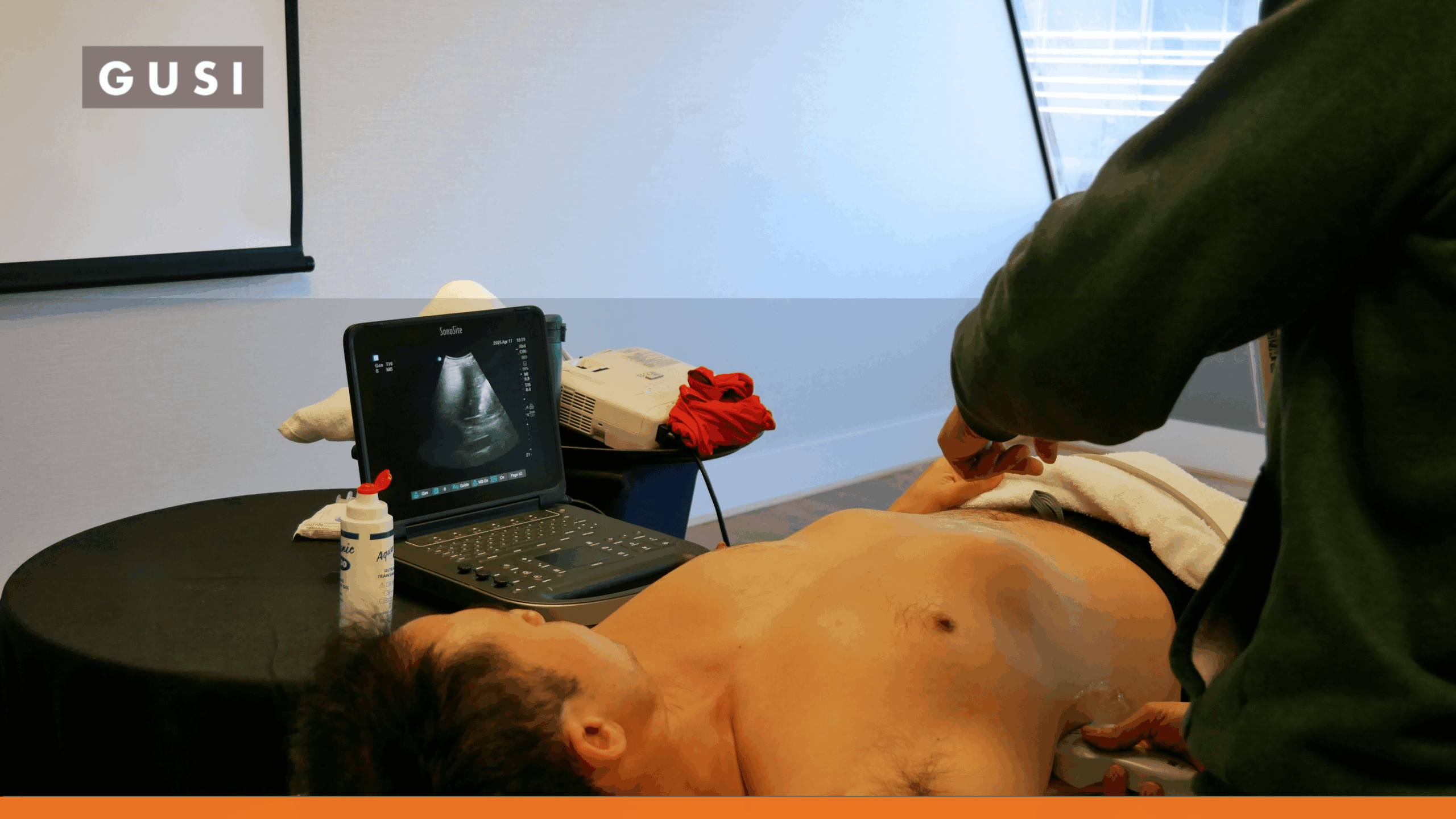


Breast complaints show up in emergency departments, primary care clinics, urgent care, postpartum care, and postoperative settings. A patient may have focal pain and redness, a fluctuant area, a new lump, nipple discharge, or concern about a surgical site. Ultrasound can be useful in many of these scenarios, but not every breast ultrasound is point-of-care ultrasound.
Breast POCUS is best thought of as a focused bedside examination performed to answer a limited clinical question. One of its clearest uses is determining whether an acutely painful or inflamed area contains a drainable fluid collection.
The Direct Answer
POCUS in breast health is most useful for focused acute questions such as whether suspected mastitis or soft-tissue infection has formed an abscess, whether a postoperative area contains a fluid collection, and where a collection lies before a selected ultrasound-guided procedure. POCUS should not replace breast cancer screening, comprehensive diagnostic breast ultrasound, mammography, BI-RADS assessment, or a formal workup of a persistent or suspicious mass.
That distinction protects patients from two opposite errors: delaying useful bedside care when a focused scan could help, and offering false reassurance when a limited scan is not enough.
What Is Breast POCUS?
POCUS is ultrasound performed and interpreted by the treating clinician as part of the current encounter. The scan begins with a focused question and should end with an action: treat, drain, refer, obtain formal imaging, or recognize that the bedside exam is nondiagnostic.
In breast care, that might mean asking whether a tender erythematous region contains a discrete fluid collection or whether a postoperative swelling appears fluid-filled. It does not mean performing an abbreviated cancer-screening examination of the entire breast.
A focused bedside scan can overlap technically with diagnostic breast ultrasound, but the scope, reporting framework, equipment requirements, personnel qualifications, and quality systems are different.
POCUS vs. Formal Diagnostic Breast Ultrasound
| Feature | Focused Breast POCUS | Formal Diagnostic Breast Ultrasound |
|---|---|---|
| Purpose | Answers a limited bedside question tied to the current encounter | Evaluates a breast symptom or imaging abnormality within a formal diagnostic pathway |
| Typical scope | Localized symptomatic area or procedure target | Targeted or comprehensive breast/axillary evaluation as clinically indicated |
| Operator | Treating clinician with appropriate POCUS training and local scope | Breast sonographer, radiologist, surgeon, or other appropriately qualified professional within a breast-imaging workflow |
| Reporting | Focused clinical note documenting question, findings, limitations, images, and next step | Formal breast imaging report using standardized terminology and management recommendations, commonly BI-RADS |
| Cancer screening | Not a substitute for screening mammography or a formal screening program | Supplemental or diagnostic ultrasound may be used in defined risk/imaging scenarios |
| Best fit | Abscess/fluid question, selected superficial procedure guidance, triage | Palpable mass, nipple discharge, focal pain, suspicious imaging finding, high-risk or supplemental imaging, biopsy planning |
The American College of Radiology (ACR) practice parameter for diagnostic breast ultrasound includes dedicated expectations for examination technique, personnel, documentation, equipment, and quality control. BI-RADS provides standardized language and management recommendations across breast imaging. [1][2]
A clinician should not present a limited bedside study as equivalent to that formal diagnostic process.
When Can POCUS Help With a Breast Complaint?
Suspected Breast Abscess
This is the strongest bedside use case. Physical examination can suggest an abscess, but inflammation, induration, edema, and tenderness do not always reveal whether a drainable cavity has formed.
POCUS can identify a fluid collection, estimate its size and depth, show internal debris or septations, and help determine whether the collection is near the skin, chest wall, nipple-areolar complex, implant, or other structures that affect management.
A 2024 review of breast infections notes that ultrasound evaluation and minimally invasive treatment have become central to abscess care. [3] General soft-tissue evidence also supports POCUS for distinguishing abscess from cellulitis, although breast-specific disease requires additional attention to lactation, malignancy, implants, surgery, and recurrence. [4]
Mastitis That Is Worsening or Not Responding as Expected
Lactational mastitis is usually diagnosed clinically, and routine imaging is not required for every patient. The American Academy of Family Physicians (AAFP’s) 2024 review recommends ultrasound to look for abscesses in patients who are immunocompromised or whose symptoms worsen or recur. [5]
POCUS can help answer the immediate fluid-collection question, but persistent, recurrent, or atypical inflammation may require formal breast imaging and specialist evaluation. Nonlactational inflammatory conditions can mimic malignancy, and some breast cancers can initially look inflammatory.
Postoperative Seroma, Hematoma, or Fluid Collection
After breast surgery, reconstruction, mammoplasty, biopsy, or another procedure, a localized swelling may represent a seroma, hematoma, infection, or another complication.
A focused scan can show whether the area is fluid-filled and whether a collection is simple or complex. It can also help determine the depth before aspiration. However, postoperative anatomy can be altered, and implant-related or persistent abnormalities often need formal imaging or surgical follow-up.
Selected Superficial Procedural Guidance
Ultrasound guidance can help a trained clinician place a needle into a visible fluid collection while avoiding adjacent structures. This may be used for aspiration or catheter placement in selected cases and within appropriate institutional and specialty workflows.
Breast abscess literature supports percutaneous ultrasound-guided aspiration as a minimally invasive treatment option, often with good cosmetic and recovery advantages, though repeated aspiration or surgical drainage may still be required depending on size, loculation, recurrence, and clinical context. [6][7]
How to Perform a Focused Breast POCUS Exam
A bedside breast exam should stay focused on the symptomatic area rather than imitating a whole-breast diagnostic study.
- Use a high-frequency linear transducer for superficial breast tissue and the skin/soft-tissue interface.
- Position the patient so that the symptomatic area is accessible and the tissue is supported. Supine or slight oblique positioning may help for lateral lesions.
- Scan directly over and around the point of maximal tenderness, swelling, erythema, palpable abnormality, or postoperative concern.
- Image the area in at least two orthogonal planes and sweep beyond the apparent margins.
- For a suspected collection, assess size in three dimensions, depth from skin, internal echoes, septations, posterior enhancement, surrounding edema, and relation to the nipple, chest wall, implant, or surgical scar.
- Color Doppler may help demonstrate surrounding hyperemia, but Doppler findings are not specific for infection or malignancy.
- Save representative images or clips and document exactly what question the exam was intended to answer.
- If no adequate window is obtained or the clinical finding is not explained, document the study as limited or nondiagnostic and escalate appropriately.
Technique Guardrail
Do not use a limited symptomatic-area scan to declare the rest of the breast normal. POCUS is only as complete as the clinical question and the area examined.
What Does a Breast Abscess Look Like on Ultrasound?
Breast abscesses do not all look like perfectly black, round cysts. The sonographic appearance can vary with timing, contents, prior treatment, and surrounding inflammation.
| Feature | Possible Appearance | Clinical Meaning |
|---|---|---|
| Fluid collection | Hypoechoic or anechoic cavity, often irregular | Supports a drainable collection when correlated clinically. |
| Internal debris | Low-level echoes, swirling material, complex fluid | Common in pus or blood; not specific by itself. |
| Posterior enhancement | Increased through-transmission behind fluid | Supports fluid content. |
| Septations / loculations | Internal linear structures or multiple pockets | May make simple aspiration less successful and alter drainage planning. |
| Surrounding edema | Cobblestoning or thickened subcutaneous tissue | Supports inflammatory change but can occur without abscess. |
| Hyperemia | Increased peripheral or surrounding Doppler flow | Can accompany active inflammation; not diagnostic of infection alone. |
| Gas / shadowing | Dirty shadowing or echogenic foci in selected infections/procedural contexts | May complicate visualization and should raise concern in the appropriate clinical setting. |
Abscess, necrotic tumor, hematoma, postoperative collection, and complicated cyst can sometimes overlap sonographically. The patient’s history and follow-up pathway matter.
Mastitis, Cellulitis, and Abscess: What POCUS Can Add
| Clinical Picture | Potential POCUS Role | What POCUS Does Not Settle |
|---|---|---|
| Early uncomplicated lactational mastitis | Often no POCUS needed if improving with appropriate care | Microbiology, cancer exclusion, or need for imaging if symptoms later persist |
| Diffuse erythema / edema without fluctuance | Look for hidden fluid collection when concern remains | The cause of inflammation |
| Focal fluctuance or severe focal tenderness | Identify and map a collection | Whether every collection should be drained at bedside |
| Worsening / recurrent symptoms | Look for an abscess or complex collection | Need for formal breast imaging, culture, biopsy, or specialist care |
| Nonlactational abscess | Identify collection and guide urgent management | Underlying duct disease, granulomatous disease, malignancy, or recurrence risk |
A New Breast Mass Is a Different Clinical Pathway
A new palpable breast mass should not be managed as a soft-tissue POCUS problem. Even if a bedside scan shows a structure that appears cystic or benign, the patient may still need formal diagnostic breast imaging based on age, pregnancy/lactation status, symptoms, and risk.
ACR guidance reflects that difference. For a palpable mass in a patient younger than 30, formal breast ultrasound is usually the initial imaging test. From ages 30 to 39, ultrasound, diagnostic mammography, or Digital Breast Tomosynthesis (DBT) may be used depending on the scenario. At age 40 and older, diagnostic mammography or DBT is usually first-line, with ultrasound added as indicated. [8]
Do Not Let ‘Looks Benign’ End the Workup
Features such as oval shape, circumscribed margins, parallel orientation, or posterior enhancement can occur in benign lesions, but morphology alone does not turn a bedside scan into a formal cancer exclusion test. Persistent or clinically concerning masses belong in the diagnostic breast-imaging pathway.
When Formal Breast Imaging Is Needed
| Presentation | POCUS Role | Formal Pathway |
|---|---|---|
| New palpable mass | May help triage an acute fluid question, but should not be the endpoint | Formal diagnostic breast imaging according to age, risk, and pregnancy/lactation status |
| Persistent focal noncyclical pain | Limited role unless looking for acute collection | ACR supports formal breast US and/or diagnostic mammography/DBT depending on age |
| Pathologic nipple discharge | POCUS is not a substitute for duct/breast evaluation | Formal diagnostic breast imaging is usually appropriate in age- and pregnancy-specific pathways |
| Nipple retraction, skin dimpling, peau d’orange, unexplained adenopathy | Do not use negative POCUS for reassurance | Prompt diagnostic breast imaging and appropriate breast specialist referral |
| Recurrent or nonlactational abscess | Useful for identifying/draining collection | Formal follow-up may be needed to evaluate underlying pathology |
| Pregnant or lactating patient with focal mass/pain | Focused POCUS may identify an obvious collection | Formal breast ultrasound is commonly appropriate initial imaging, with mammography/DBT added in selected patients |
| Implant concern | May reveal peri-implant fluid in some situations | Use implant-specific formal imaging pathway; modality depends on implant type, age, symptoms, and suspected complication |
Can POCUS Screen for Breast Cancer?
No. A focused bedside breast scan should not be used as a substitute for recommended breast cancer screening.
The United States Preventive Services Task Force (USPSTF) currently recommends biennial screening mammography for average-risk women from age 40 through 74. Other organizations, including the ACR, recommend different schedules and emphasize earlier risk assessment for patients who may need intensified screening. [9][10]
Those recommendations concern organized screening and dedicated breast-imaging modalities. POCUS is not a validated replacement for mammography, DBT, screening Magnetic Resonance Imaging (MRI), or a formal supplemental whole-breast ultrasound program.
Breast Density and Supplemental Ultrasound
Dense breast tissue can reduce mammographic sensitivity and is itself a breast cancer risk factor, but supplemental imaging should be individualized.
ACR’s 2024 Appropriateness Criteria state that supplemental breast ultrasound may be appropriate in some dense-breast and elevated-risk scenarios, while MRI is usually preferred in several high-risk or extremely dense categories. [11]
That is very different from saying every patient with dense breasts should receive a quick bedside POCUS screen. Supplemental breast ultrasound is a formal breast-imaging examination performed within a screening or diagnostic program.
Ultrasound-Guided Aspiration and Drainage
When a confirmed fluid collection is appropriate for percutaneous treatment, ultrasound guidance can improve localization and help avoid blind aspiration.
Current breast-abscess literature supports ultrasound-guided needle aspiration as a minimally invasive option in many patients, with potential benefits in healing time, cosmesis, and continued breastfeeding. Larger, multiloculated, recurrent, postoperative, or treatment-resistant collections may require repeated aspiration, catheter drainage, or surgery. [6][7]
The clinician must also decide what material needs culture or other testing and whether the patient needs breast surgery, radiology, lactation, infectious disease, plastic surgery, or oncology follow-up.
This article does not prescribe an antibiotic regimen because treatment depends on context and local microbiology.
Common Pitfalls and Safety Risks
- Using POCUS as a breast cancer screening examination.
- Calling a mass benign because it looks oval, circumscribed, or cystic on a limited scan without the correct diagnostic pathway.
- Assuming a negative POCUS examination excludes malignancy or a deep collection.
- Scanning only the center of a painful area and missing an eccentric or deeper collection.
- Confusing postoperative hematoma, seroma, necrotic mass, complicated cyst, or implant-related fluid with an uncomplicated abscess.
- Draining a lesion without confirming the clinical context, procedural scope, anticoagulation status, nearby implant, anatomy, and follow-up plan.
- Treating recurrent nonlactational abscesses as repeated simple infections without considering underlying pathology.
- Failing to escalate nipple retraction, pathologic discharge, suspicious skin change, unexplained adenopathy, or a persistent mass.
- Using Breast Imaging Reporting and Data System (BI-RADS) terminology casually without the training, complete examination, reporting structure, and follow-up system that BI-RADS is designed to support.
Training, Documentation, and Quality Assurance
Breast complaints are emotionally charged and clinically high-stakes because acute infection and malignancy can overlap. Training therefore needs to cover more than finding fluid.
Clinicians using POCUS for breast complaints should understand superficial ultrasound technique, abscess versus cellulitis, normal breast and chest-wall anatomy, postoperative changes, procedural guidance, infection-control practices, limitations, documentation, and the formal breast-imaging referral pathway.
GUSI’s POCUS Essentials curriculum includes skin and soft-tissue scanning for cellulitis versus abscess, which provides a relevant technical foundation. Breast-specific use still requires attention to dedicated breast-imaging standards and local clinical governance. [12]
Representative images, lesion location, measurements, depth, limitations, procedural images when relevant, and the referral/follow-up plan should be documented according to local policy.
Training Principle
The goal is not to teach a frontline clinician to replace the breast imaging service. It is to help the clinician answer an urgent focused question safely, recognize when the answer is incomplete, and route the patient into the right next step.
Case: Postoperative Breast Abscess
A 28-year-old woman with a history of bilateral mammoplasty presented with acute right-breast tenderness, erythema, edema, warmth, and low-grade fever. Examination showed focal fluctuance. Bedside ultrasound identified a developing collection measuring approximately 1.9 x 1.0 x 1.2 cm.
Ultrasound guidance was used to aspirate the collection, yielding sanguinopurulent material for culture. Follow-up ultrasound showed interval decrease in the collection, and the patient was referred back to her surgical team.
The case illustrates a good POCUS use: the clinician had a focused question, identified a superficial collection, used ultrasound to guide a procedure, sent appropriate diagnostic material, and arranged follow-up.
It also illustrates why POCUS was not the entire breast-health evaluation. The patient had prior surgery, recurrent local symptoms, and ultimately required ongoing surgical management. The bedside scan answered the acute fluid question; it did not replace the broader postoperative assessment.