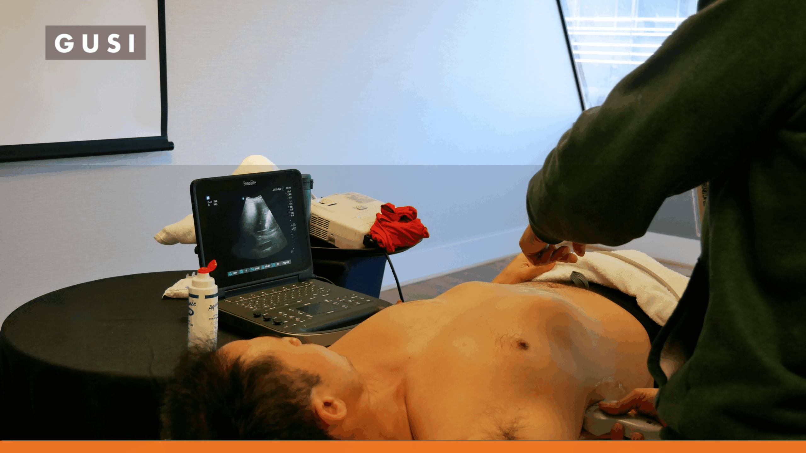




Point-of-care ultrasound (POCUS) is increasingly becoming part of the primary care toolkit. Instead of sending every focused imaging question outside the clinic, a trained family physician or general practitioner can use ultrasound during the patient encounter to look for specific findings that may clarify the next step.
That does not turn a primary care visit into a radiology department. POCUS is a focused examination performed to answer a defined clinical question and interpreted in the context of the history, physical examination, and the clinician’s scope of practice.
The Direct Answer
POCUS in primary care is the focused use of ultrasound by a trained clinician during the patient encounter to support a specific diagnostic or procedural question. It can help speed clinical decisions, reduce some referrals, and improve access to imaging, but it does not replace comprehensive diagnostic ultrasound or other imaging when a broader evaluation is needed.
What Is POCUS in Primary Care?
A comprehensive diagnostic ultrasound is generally performed as a formal imaging study with a defined protocol, extensive image acquisition, and interpretation by an imaging specialist or other appropriately credentialed clinician. POCUS is narrower by design.
The primary care clinician starts with a clinical question: Is there a full bladder? Is hydronephrosis present? Is there an abdominal aortic aneurysm? Are lung findings consistent with pulmonary edema? Is there a joint effusion? Is the fetal presentation cephalic or breech in a clinician whose practice includes obstetric POCUS?
The scan is used alongside the rest of the examination to refine probability, guide a procedure, determine whether more testing is needed, or decide how urgently a patient should be referred.
Why POCUS Fits the Primary Care Visit
Primary care is built around undifferentiated symptoms, continuity, and deciding what needs to happen next. That makes the point-of-care model particularly relevant. The value is not that ultrasound can answer every question. It is that a limited number of well-chosen scans can sometimes provide useful information while the clinician and patient are still in the room.
| Primary Care Need | Potential Role for POCUS |
|---|---|
| Clarify a focused question | Adds real-time imaging to the history and physical examination. |
| Reduce delays | Provides an immediate focused assessment instead of waiting for external imaging when appropriate. |
| Improve referral quality | Helps determine whether a patient can be managed in a clinic or needs formal imaging or specialist evaluation. |
| Support rural or resource-limited care | Brings imaging capability closer to the patient when access to radiology is limited. |
| Guide procedures | Visualizes anatomy during selected injections, drainage, vascular access, or other procedures. |
| Reassess over time | Repeats focused examinations without ionizing radiation when serial assessment is clinically appropriate. |
World Organization of Family Doctors (WONCA) Europe’s 2024 position paper states that POCUS has an increasingly important role for specific primary care indications and recommends that GPs receive training tailored to their healthcare context. The paper also highlights portability, patient acceptability, diagnostic confidence, potential reductions in referrals, and the importance of ongoing competency and risk management. [1]
What the Evidence Shows
WONCA Europe Endorses POCUS in Family Medicine
WONCA Europe represents family medicine organizations across Europe. Its position statement endorses POCUS within family medicine and recommends curriculum-based training during residency and continuing medical education. It also stresses that indications vary by country, healthcare system, local scope, and clinician training. [1]
That nuance matters. The case for POCUS in primary care is not “every GP should perform every scan.” It is that appropriately trained clinicians can use selected applications that match the needs of their patients and practice environment.
Real-World Use Can Change Primary Care Decisions
One frequently cited prospective study followed 20 experienced Danish GPs who used POCUS in everyday office practice. Across 574 patients, the median scan took five minutes. POCUS changed the main tentative diagnosis in 49.4% of consultations, changed the management plan in 50.9%, and changed treatment in 26.5%. Intended referrals to secondary care fell from 49.2% before POCUS to 25.6% after the scan. [2]
These numbers show that POCUS can materially influence decision-making, but they should not be interpreted as proof that every change was better. The study involved clinicians who already used ultrasound regularly, and the authors explicitly noted that they did not determine whether each change improved patient outcomes.
Newer Research Strengthens the Case for Structured Training
Primary care ultrasound is highly operator-dependent, which is why recent research has shifted toward competency and implementation rather than simply device access.
In a 2025 study, 18 office-based GPs completed a three-month training program tailored to primary care. Their measured scanning competence ranged from 68.9% to 82.3% of the maximum score at course completion and improved to 80.9% to 92.6% at three-month follow-up across the applications studied. [4]
A separate 2025 follow-up of 564 patients scanned in general practice found a low but real risk of possible misdiagnosis, potential overdiagnosis, and incidental findings. The authors concluded that GP-performed POCUS was generally safe while still capable of causing unnecessary testing or harm in some cases. [5]
In 2026, European Federation of Societies for Ultrasound in Medicine and Biology (EFSUMB) published a primary-care POCUS core curriculum and training recommendations. A related European Delphi process involving 95 experienced primary care physicians from 28 countries identified 40 diagnostic examinations across 13 anatomical areas for a basic core curriculum. [6][7]
What the Evidence Means
POCUS can be useful in primary care, but the device is the easy part. Safe adoption depends on selecting the right applications, building competency, maintaining skills, documenting findings, reviewing image quality, and knowing when a focused scan is not enough.
Common POCUS Applications in Primary Care
The right application depends on the clinician’s training, patient population, local regulations, and practice scope. Current American Academy of Family Physicians (AAFP) training for family medicine includes applications such as skin and soft tissue, musculoskeletal ultrasound, abdominal aortic aneurysm screening, basic cardiac and lung assessment, renal and bladder ultrasound, and selected pregnancy-related examinations. [8][9]
| Clinical Question | How POCUS May Help | When to Escalate |
|---|---|---|
| Dyspnea or edema | Focused lung and cardiac views may identify findings such as B-lines, pleural fluid, pericardial effusion, or gross ventricular dysfunction. | Uncertain findings, suspected pulmonary embolism, ischemia, complex heart disease, or clinical instability requiring definitive evaluation. |
| Possible abdominal aortic aneurysm | Measure the abdominal aorta in appropriately selected patients. | Symptoms concerning for rupture, technically limited exam, abnormal measurement, or need for formal vascular assessment. |
| Flank pain / urinary symptoms | Assess for hydronephrosis and bladder distention or retention. | Atypical or severe presentation, infection/obstruction concerns, renal dysfunction, uncertain diagnosis, or need for stone characterization. |
| Right upper quadrant pain | Look for gallstones and selected sonographic features associated with biliary disease. | Equivocal scan, suspected complications, alternative diagnosis, jaundice, systemic illness, or persistent concerning symptoms. |
| Joint or soft-tissue complaint | Identify joint effusion, tendon abnormalities, abscess, foreign body, or other selected superficial findings. | Deep infection, major trauma, uncertain anatomy, suspected malignancy, or pathology beyond the operator’s validated scope. |
| Pregnancy-related focused question | Within appropriate obstetric scope, assess selected questions such as fetal cardiac activity, presentation, or early pregnancy landmarks. | Pain/bleeding with concern for ectopic pregnancy, uncertain viability/location, maternal instability, abnormal findings, or need for comprehensive obstetric imaging. |
| Procedure guidance | Visualize anatomy during selected injections, drainage, vascular access, or other procedures. | Poor visualization, anatomy outside clinician competence, complication risk, or procedure requiring specialist care. |
Lung and Dyspnea
Lung ultrasound can add useful information when a primary care patient presents with shortness of breath, cough, or suspected fluid overload. A focused scan may identify B-lines, pleural effusion, consolidation patterns, or absence of normal lung sliding. These findings are not interpreted in isolation. They modify the clinical assessment and may help determine whether the patient can be managed locally or needs chest radiography, CT scan, echocardiography, emergency evaluation, or specialist care.
Focused Cardiac Assessment
A limited cardiac exam can answer narrower questions than a comprehensive echocardiogram. Depending on training, primary care clinicians may assess gross ventricular function, pericardial fluid, or selected volume-status findings. POCUS should not be used to claim detailed valvular, structural, or hemodynamic diagnoses that require comprehensive echocardiography.
Abdominal Aortic Aneurysm
Ultrasound is well suited to focused aortic measurement, making abdominal aortic aneurysm screening and selected symptomatic assessments a practical primary care application. A positive, incomplete, or clinically concerning scan requires appropriate confirmatory imaging and referral.
Renal and Bladder Questions
POCUS can help evaluate bladder volume, urinary retention, and hydronephrosis. It may be particularly helpful when the question is whether obstruction is plausible or whether bladder distention explains the patient’s symptoms. It does not identify every cause of flank or abdominal pain, and a negative focused scan does not rule out nephrolithiasis or other important pathology.
Gallbladder and Focused Abdominal Questions
In clinicians trained for the application, POCUS can assess gallstones and selected gallbladder findings. The focused scan can be useful when it matches the clinical picture, but an incomplete or discordant examination should lead to comprehensive imaging or further evaluation rather than false reassurance.
Musculoskeletal and Soft Tissue
Primary care and sports medicine clinicians may use ultrasound to assess superficial tendons, joint effusions, abscesses, foreign bodies, and selected soft-tissue complaints. Ultrasound guidance can also improve visualization during procedures. Training should be application-specific because normal anatomy, probe handling, and interpretation differ substantially across body regions.
Pregnancy and Obstetric Care Within Scope
Family physicians whose scope includes obstetric care may use focused ultrasound for selected pregnancy-related questions. The distinction between a limited bedside scan and a comprehensive obstetric ultrasound must remain explicit. Local privileges, reporting standards, and referral pathways are especially important.
POCUS vs. Comprehensive Diagnostic Ultrasound
| Feature | POCUS | Comprehensive Diagnostic Ultrasound |
|---|---|---|
| Clinical purpose | Answer a focused question tied to the current encounter | Evaluate an organ/system using a formal imaging protocol |
| Operator | Treating clinician with validated training for the application | Sonographer, radiologist, cardiologist, obstetric imaging specialist, or other appropriately credentialed professional |
| Scope | Limited and question-specific | Broader and protocol-driven |
| Timing | During the visit or bedside encounter | Scheduled or performed through an imaging service |
| Interpretation | Integrated immediately with the clinical assessment | Formal interpretation and report |
| Best use | Focused decision support and procedural guidance | Comprehensive anatomic evaluation, definitive imaging, or specialist-level detail |
The modalities are complementary. A well-performed POCUS exam can answer the first question and make the next step clearer. It should not be stretched beyond its validated purpose simply to avoid a referral.
When POCUS Should Not Be the Final Test
The safest POCUS programs teach clinicians not only how to scan, but also when to stop scanning and escalate. A focused examination should not delay comprehensive imaging or specialist evaluation when:
- The patient is unstable or has a time-sensitive emergency that requires definitive imaging or treatment.
- The scan is technically limited, nondiagnostic, or outside the clinician’s validated application.
- A negative POCUS result does not adequately reduce the probability of serious disease.
- The symptoms and ultrasound findings do not fit together.
- The condition requires detailed anatomic mapping, staging, procedural planning, or formal measurement.
- An incidental or unexpected finding needs confirmation.
- Local policy, scope of practice, credentialing, or reimbursement rules require formal imaging.
Training, Competency, and Quality Assurance
Buying an ultrasound machine does not create a POCUS program. Competence combines knowledge of anatomy and pathology, image acquisition, image optimization, interpretation, documentation, and clinical decision-making.
WONCA Europe recommends stepwise, ongoing training and continuing education. EFSUMB’s 2026 recommendations similarly emphasize a standardized foundation that can be adapted to local practice. In the United States, the AAFP maintains family medicine POCUS curriculum and training resources. [1][6][8][9]
| Program Component | What Good Implementation Looks Like |
|---|---|
| Application scope | Start with a limited group of high-value scans matched to the practice’s patient population. |
| Foundational education | Ultrasound physics, knobology, anatomy, normal findings, pathology, artifacts, and limitations. |
| Hands-on practice | Repeated supervised scanning rather than lecture-only learning. |
| Competency assessment | Demonstrate acquisition, interpretation, documentation, and clinical integration for each application. |
| Image archiving | Store representative images/clips when required for clinical care, QA, billing, or credentialing. |
| Documentation | Record indication, exam type, key findings, limitations, interpretation, and resulting plan. |
| Quality review | Use structured review and feedback to identify image-quality and interpretation errors. |
| Maintenance | Continue scanning, review edge cases, and reassess skills when an application is used infrequently. |
| Escalation | Define when formal ultrasound, CT/MRI, radiology, emergency care, or specialist consultation is required. |
How to Implement POCUS in a Primary Care Practice
- Start with the patient problems you see most often, not with a list of everything the device can scan.
- Choose one to three applications where a focused answer can realistically change the next step.
- Build a formal training pathway with supervised scanning and competency assessment.
- Create documentation, image-storage, billing, privacy, and quality-review workflows before routine clinical use.
- Define escalation criteria for negative, limited, unexpected, or discordant examinations.
- Track utilization, diagnostic impact, referral patterns, scan quality, and adverse or near-miss events.
- Expand the application set only after clinicians demonstrate and maintain competence in the initial scope.
The Goal Is Better Decisions, Not More Scanning
POCUS is most valuable when it improves a real clinical decision. A scan that does not answer a focused question, change management, guide a procedure, or improve triage may simply add time and uncertainty.
For primary care, the long-term opportunity is not to turn every visit into an ultrasound examination. It is to give clinicians another bedside tool that can shorten the path from uncertainty to an appropriate next step when the application, patient, and operator are a good match.
That is also why training and feedback matter. A confident clinician who understands both what POCUS can show and what it can miss is more useful to the patient than a clinician with access to the latest handheld device but no structured pathway for competency and review.
Build Practical POCUS Skills for Family Medicine
GUSI offers POCUS training designed for family medicine and primary care clinicians, including online learning, hands-on courses, scan review, and tools for ongoing skill development. Explore the Family Medicine training pathway to build the acquisition, interpretation, documentation, and clinical judgment needed to use POCUS responsibly.