
Opens in a new tab

Automated image analysis

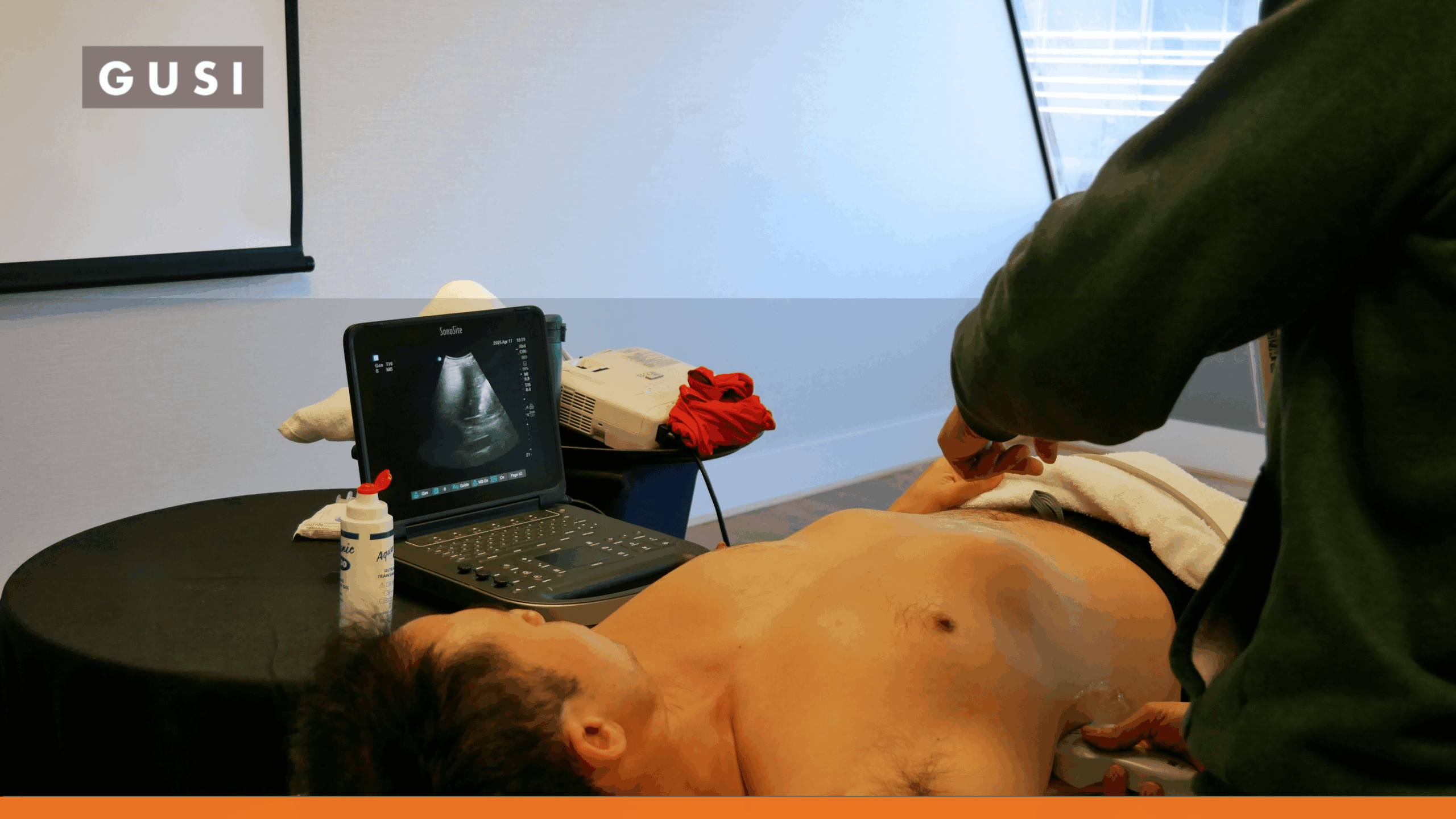
Automated image analysis in medical ultrasound streamlines educational and administrative processes by employing AI and machine learning algorithms to interpret sonographic data. This technology accelerates the learning curve for students by providing instant feedback and objective assessment of scans, reducing the need for extensive manual review by instructors. For administrators, it optimizes workflow efficiency and resource allocation by automating quality control, data indexing, and reporting, thereby enhancing the overall operational management within educational institutions and healthcare facilities.

This advanced approach to image interpretation also aids in standardizing training modules and performance evaluations. By consistently analyzing vast datasets, it identifies patterns and anomalies faster than human observation, serving as a powerful tool for both diagnostic support and continuous professional development in ultrasound. This innovation significantly improves the accuracy and speed of medical imaging workflows, fostering a more efficient and effective learning and operational environment.
More Definitions

Want to learn more?

POCUS Essentials Course

Learn More

Related Posts by Term

POCUS QA process

The QA Gap: Why Most POCUS Programs Still Don’t Have One and How to Fix It

POCUS training can give clinicians the confidence to scan. But
continue reading

How AI Will Change the Way You Practice POCUS — Insights From Our Expert Panel

Part 1 of a 3-Part Series on Artificial Intelligence in
continue reading

Building the Next Generation of Ultrasound Educators: The GUSI MedConnect Instructor Training Program

In a small practice room at Touro University California, a
continue reading

MedConnect – Bruno Vargas

“I don’t know how to be a doctor without using
continue reading

Closing the Maternal Care Gap in Rural Philippines With POCUS

Bridging the Maternal Care Gap with POCUS In many rural
continue reading

How Lung Ultrasound Helps Clinicians Catch Tuberculosis Sooner

Webinar Summary: June 18, 2025 | Presented by Global Ultrasound
continue reading

The FASH Exam: POCUS for HIV-Associated Tuberculosis

The FASH exam is a focused POCUS protocol that helps
continue reading

Hands-On POCUS Training for Primary Care in San Francisco

See how GUSI teaches hands-on POCUS for primary care in
continue reading

POCUS vs. CT Scan: When Ultrasound Is the Safer First Step

POCUS provides immediate, radiation-free answers to focused clinical questions, while
continue reading

Contribute to the POCUSpedia

Help us improve the accuracy and completeness of the POCUSpedia. Your contributions help maintain the highest standards of factual content.
Contributors who provide verified corrections or additions will be credited with a link to their chosen web address. We appreciate your expertise and time in helping us maintain accurate information about POCUS

As a [Specialty] clinician, POCUS has completely changed how I practice. GUSI’s training gave me the confidence to make faster, more accurate decisions for my patients.

JONATHAN DOE
Private Practice Owner

Suggest a Correction

* Required fields

“”