
Muscle

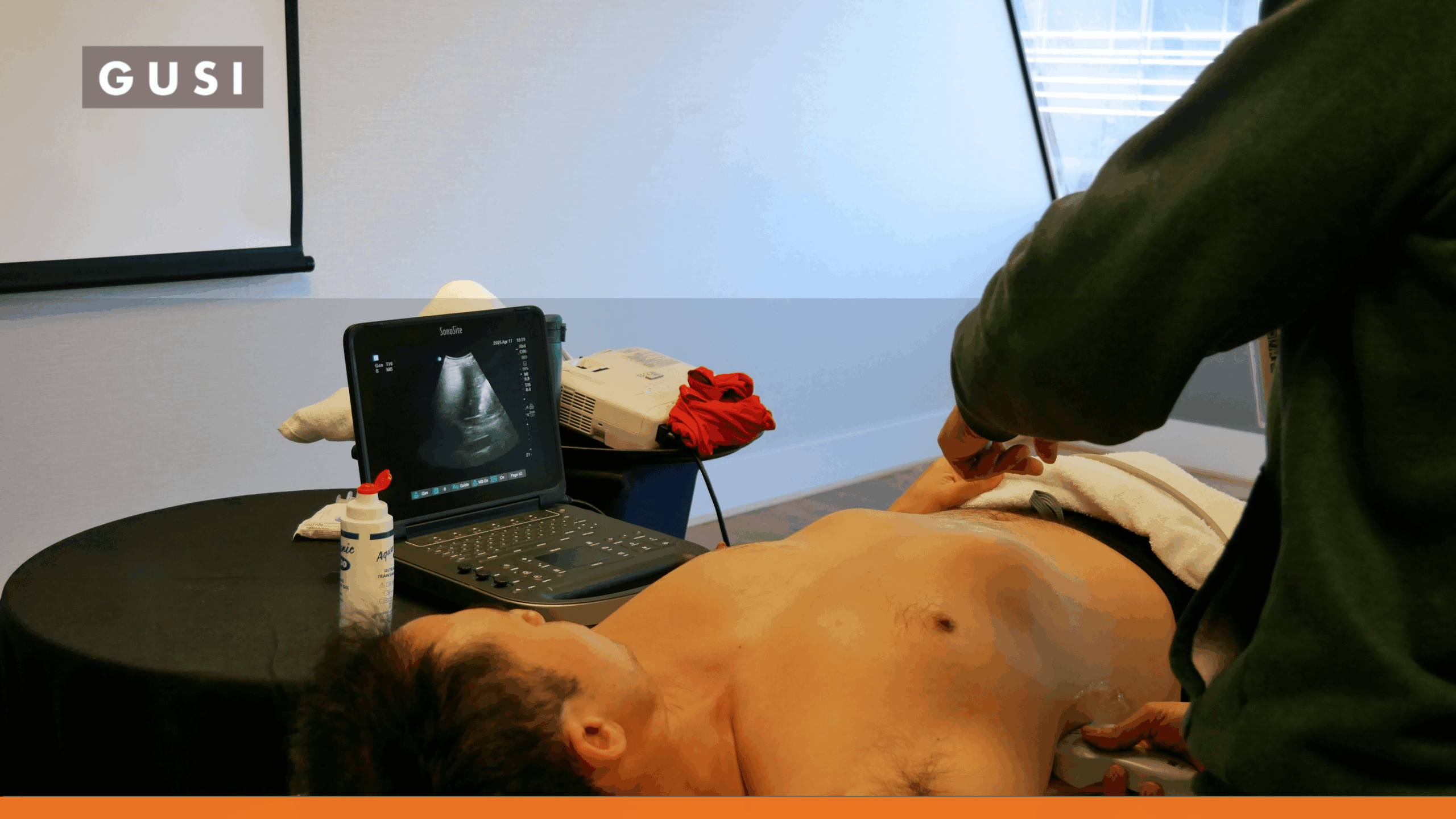
In musculoskeletal (MSK) ultrasound, muscles are critical soft tissue structures visualized for diagnostic purposes. These contractile tissues, essential for movement and posture, appear with a characteristic pennate or parallel fibrillar echotexture on ultrasound images. Understanding their normal sonographic appearance is fundamental for identifying pathology.

Medical professionals leverage MSK ultrasound to assess muscle injuries such as strains, tears, and hematomas, as well as inflammatory conditions like myositis. Ultrasound provides real-time, dynamic evaluation of muscle function, allowing for precise localization of abnormalities and guiding therapeutic interventions.


Clinical Author

Dr. Emma Khabure is an experienced Family Physician in Kenya with a dedicated focus on point-of-care ultrasound (POCUS) across clinical settings. Over the past 3 years, Dr. Emma Khabure has...

Read More
Want to learn more?

POCUS Essentials Course

Learn More

Related Posts by Term

Beginning POCUS Learner? Overcoming Barriers and Forming Habits | June 2024 Newsletter

GUSI’s pocus fellowship experience program improves access to clinical ultrasound
continue reading

Spotlight on Women’s Wellness: The Impact of POCUS in Breast Health Mar 2024 Newsletter

A firsthand account of growth and confidence through GUSI’s POCUS
continue reading

Contribute to the POCUSpedia

Help us improve the accuracy and completeness of the POCUSpedia. Your contributions help maintain the highest standards of factual content.
Contributors who provide verified corrections or additions will be credited with a link to their chosen web address. We appreciate your expertise and time in helping us maintain accurate information about POCUS

As a [Specialty] clinician, POCUS has completely changed how I practice. GUSI’s training gave me the confidence to make faster, more accurate decisions for my patients.

JONATHAN DOE
Private Practice Owner

Suggest a Correction

* Required fields

“”