
Retrovesical space

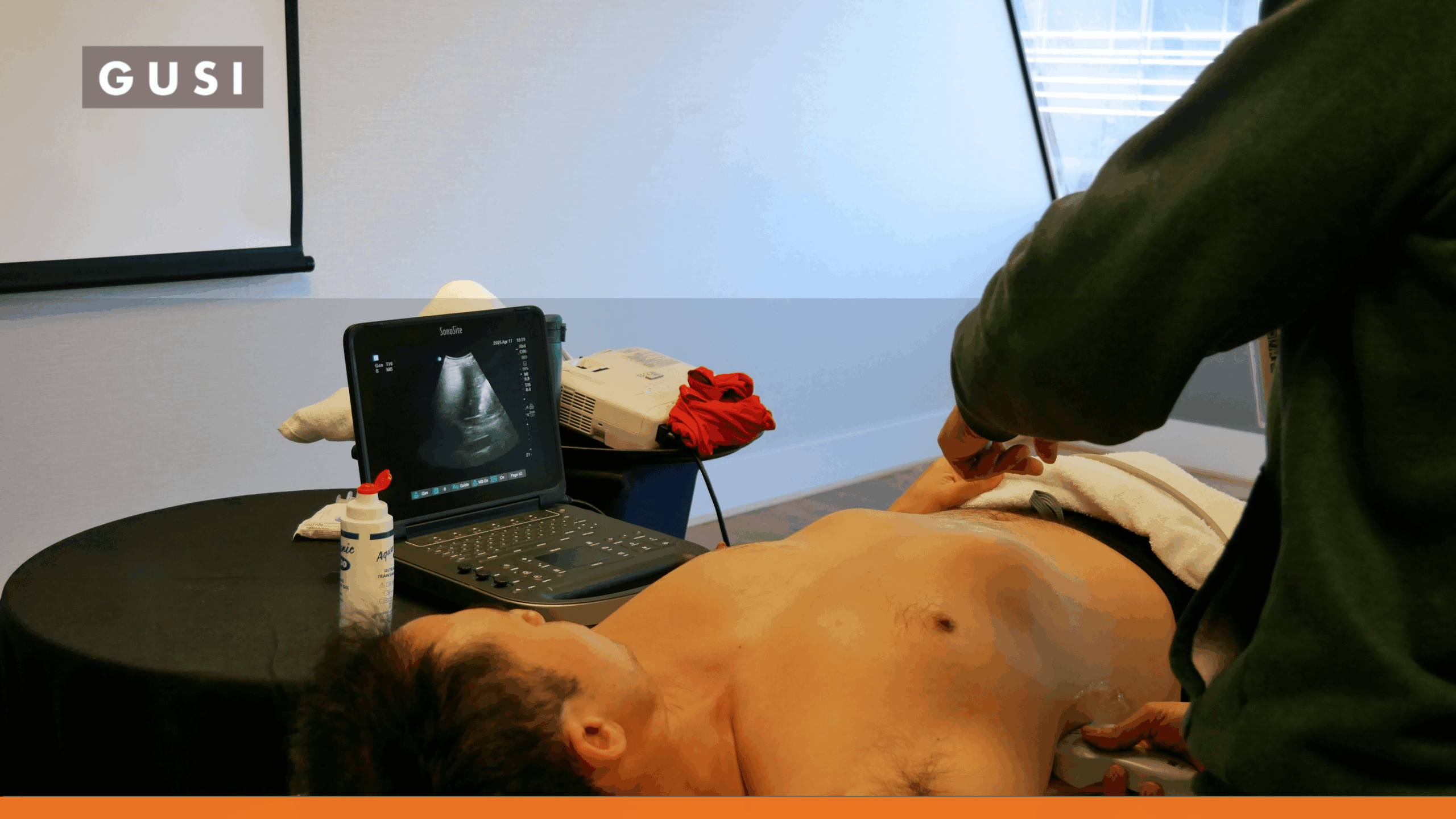
The retrovesical space in FAST/Trauma ultrasound refers to the potential space located behind the urinary bladder and in front of the rectum in males, or behind the bladder and in front of the uterus/vagina in females. This anatomical region is crucial for identifying free fluid, such as blood or urine, that may accumulate following blunt or penetrating abdominal trauma. Its visualization during a FAST (Focused Assessment with Sonography for Trauma) exam can indicate significant internal injuries.

Recognizing fluid in the retrovesical space is a key diagnostic finding in medical ultrasound, guiding rapid management decisions in trauma patients. Early detection of fluid in this area can prompt further investigation and intervention, significantly impacting patient outcomes. This ultrasound view provides critical insights into potential pelvic or abdominal hemorrhage, making it an essential component of emergency sonography protocols.


Clinical Author

Dr. Emma Khabure is an experienced Family Physician in Kenya with a dedicated focus on point-of-care ultrasound (POCUS) across clinical settings. Over the past 3 years, Dr. Emma Khabure has...

Read More
Want to learn more?

POCUS Essentials Course

Learn More

Related Posts by Term

POCUS QA process

The QA Gap: Why Most POCUS Programs Still Don’t Have One and How to Fix It

POCUS training can give clinicians the confidence to scan. But
continue reading

How AI Will Change the Way You Practice POCUS — Insights From Our Expert Panel

Part 1 of a 3-Part Series on Artificial Intelligence in
continue reading

Building the Next Generation of Ultrasound Educators: The GUSI MedConnect Instructor Training Program

In a small practice room at Touro University California, a
continue reading

MedConnect – Bruno Vargas

“I don’t know how to be a doctor without using
continue reading

Closing the Maternal Care Gap in Rural Philippines With POCUS

Bridging the Maternal Care Gap with POCUS In many rural
continue reading

How Lung Ultrasound Helps Clinicians Catch Tuberculosis Sooner

Webinar Summary: June 18, 2025 | Presented by Global Ultrasound
continue reading

The FASH Exam: POCUS for HIV-Associated Tuberculosis

The FASH exam is a focused POCUS protocol that helps
continue reading

Hands-On POCUS Training for Primary Care in San Francisco

See how GUSI teaches hands-on POCUS for primary care in
continue reading

POCUS vs. CT Scan: When Ultrasound Is the Safer First Step

POCUS provides immediate, radiation-free answers to focused clinical questions, while
continue reading

Contribute to the POCUSpedia

Help us improve the accuracy and completeness of the POCUSpedia. Your contributions help maintain the highest standards of factual content.
Contributors who provide verified corrections or additions will be credited with a link to their chosen web address. We appreciate your expertise and time in helping us maintain accurate information about POCUS

As a [Specialty] clinician, POCUS has completely changed how I practice. GUSI’s training gave me the confidence to make faster, more accurate decisions for my patients.

JONATHAN DOE
Private Practice Owner

Suggest a Correction

* Required fields

“”