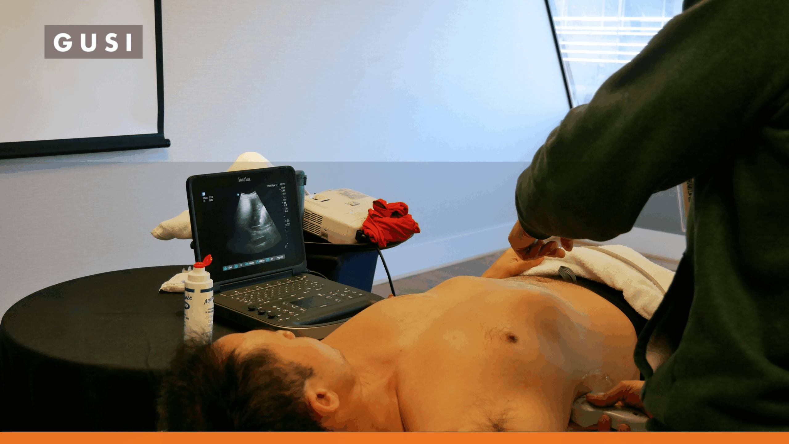

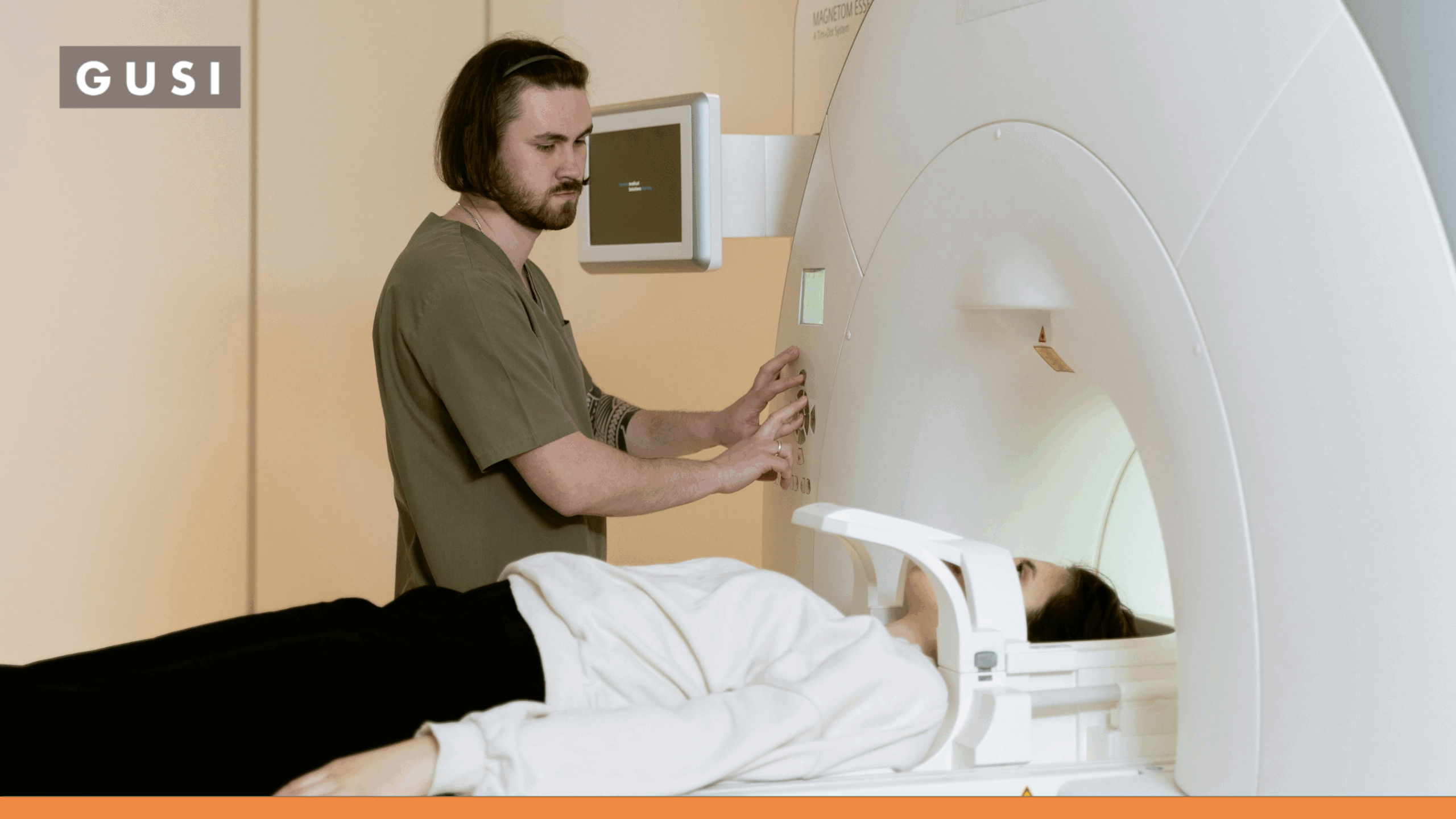

Computed tomography (CT) is one of modern medicine’s most valuable diagnostic tools. It can reveal internal bleeding, complex injuries, tumors, vascular emergencies, and other conditions that may be difficult or impossible to evaluate with ultrasound alone. But CT also uses ionizing radiation, and the patient may need to leave the bedside or clinical area to complete the scan.
Point-of-care ultrasound (POCUS) offers a different advantage. It uses sound waves rather than ionizing radiation and can provide immediate information while the clinician is examining the patient. For focused clinical questions, that combination can make POCUS a safer and faster first imaging step.
The direct answer
POCUS is safer than CT from a radiation standpoint because ultrasound does not use ionizing radiation. However, POCUS is not a universal replacement for CT. The right test depends on the clinical question, the patient’s condition, the operator’s skill, and whether a focused bedside answer is sufficient or comprehensive imaging is required.
The most useful comparison is therefore not “Which test is better?” It is “Which test can answer the question safely, accurately, and without delaying the care this patient needs?”
POCUS vs. CT Scan at a Glance
|
Comparison |
POCUS |
CT scan |
|---|---|---|
|
How images are created |
High-frequency sound waves |
Multiple x-ray measurements reconstructed into cross-sectional images |
|
Ionizing radiation |
None |
Yes |
|
Location |
At the bedside or point of care |
Usually in a dedicated imaging area |
|
Speed |
Can answer a focused question immediately |
Often rapid once performed, but may require transport, scheduling, contrast, and image interpretation |
|
Field of view |
Focused and question-specific |
Broad and comprehensive |
|
Anatomic detail |
Strong for fluid, motion, superficial structures, and selected organs |
High-detail evaluation of deep, complex, and overlapping anatomy |
|
Repeat examinations |
Can be repeated frequently without radiation exposure |
Repeated exams add cumulative radiation exposure |
|
Main limitation |
Operator-dependent; image quality may be limited by body habitus, bowel gas, dressings, wounds, or anatomy |
Radiation, possible contrast-related considerations, transport, cost, and availability |
|
Best role |
Rapid bedside assessment and serial reassessment |
Definitive or comprehensive cross-sectional evaluation |
How POCUS and CT Create Images
POCUS Uses Sound Waves at the Bedside
POCUS is a focused ultrasound examination performed and interpreted by the treating clinician at the point of care. The probe sends sound waves into the body and receives returning echoes, which are translated into real-time images. Because ultrasound uses sound rather than x-rays, it does not expose the patient to ionizing radiation. [1]
The real-time nature of POCUS is especially valuable when the clinical question involves movement or changing physiology, such as cardiac activity, fluid status, lung sliding, fetal cardiac activity, or the response to treatment. The clinician can integrate the images immediately with the history, physical examination, and hemodynamic status.
CT Uses X-Rays to Create Detailed Cross-Sectional Images
A CT scanner rotates an x-ray source around the patient and reconstructs the measurements into detailed cross-sectional images. CT can evaluate a much larger field of view than a focused POCUS exam and is often substantially better for deep structures, complex anatomy, subtle injuries, intracranial disease, and conditions that require comprehensive mapping.
That diagnostic power is why CT should never be withheld when it is the appropriate test. The safety goal is not to avoid CT. It is to use CT when its expected benefit outweighs its risks and to consider radiation-free alternatives when they can answer the clinical question without compromising care.
Radiation: The Most Important Safety Difference
Ultrasound does not use ionizing radiation. CT does. The radiation dose from a CT examination varies significantly based on the body region, patient size, scan protocol, equipment, and whether the study includes multiple phases. The U.S. Food and Drug Administration notes that estimates for diagnostic CT procedures commonly fall within about 1 to 10 millisieverts (mSv), although an individual examination may be higher or lower. [2]
Typical Adult CT Radiation Doses
|
CT examination |
Typical effective dose |
Approximate background-radiation equivalent |
|---|---|---|
|
Brain CT |
1.6 mSv |
About 7 months |
|
Chest CT |
6.1 mSv |
About 2 years |
|
Abdomen and pelvis CT |
7.7 mSv |
About 2.6 years |
|
Abdomen and pelvis CT, repeated with and without contrast |
15.4 mSv |
About 5.1 years |
|
Coronary CT angiography |
8.7 mSv |
About 3 years |
Typical values for an average-sized adult from RadiologyInfo.org, reviewed April 15, 2025. Actual dose can vary substantially and should not be used to calculate an individual patient’s exact risk. [3]
Radiation risk is cumulative, which makes unnecessary repeat imaging an important concern. A 2025 risk-modeling study in JAMA Internal Medicine projected that the 93 million CT examinations performed in the United States in 2023 could contribute to approximately 103,000 future cancers over the lifetimes of the exposed patients. The authors emphasized CT justification and dose optimization. [4]
Important context
The JAMA estimate is a population-level projection based on radiation-risk models. It does not mean that a specific CT scan will cause cancer, and it should not discourage a patient from receiving medically necessary imaging. The immediate benefit of a properly indicated CT can greatly exceed its small long-term radiation risk.
When POCUS May Be the Better First Test
POCUS is most useful when the clinician needs an immediate answer to a focused, actionable question. In those situations, bedside ultrasound may reduce delays, avoid transport, support serial reassessment, and sometimes reduce the need for CT or other imaging.
Unstable or Critically Ill Patients
A clinician can use POCUS during the initial assessment of shock, respiratory distress, trauma, or cardiac arrest to look for a limited set of urgent findings. Examples include pericardial effusion, impaired cardiac function, abdominal free fluid, pneumothorax, pleural fluid, or a markedly enlarged abdominal aorta.
This does not mean a negative POCUS exam rules out serious disease. In trauma, for example, a focused ultrasound examination can identify free fluid or thoracic findings quickly, but a stable patient may still require CT to evaluate injuries that ultrasound cannot reliably detect. A technically limited or negative study should never create false reassurance when the clinical picture remains concerning.
Focused Gallbladder and Biliary Questions
Ultrasound is commonly an appropriate initial test for right upper quadrant pain and suspected biliary disease. The American College of Radiology rates abdominal ultrasound as “usually appropriate” for initial imaging in suspected biliary disease, while CT may be used when ultrasound is negative, equivocal, or a complication or alternative diagnosis is suspected. [5]
POCUS can help identify gallstones, gallbladder wall changes, pericholecystic fluid, biliary dilation, and a sonographic Murphy sign. The findings must still be interpreted within the full clinical picture, and comprehensive imaging may be needed when the examination is incomplete or the diagnosis remains uncertain.
Selected Renal-Colic and Hydronephrosis Evaluations
POCUS can assess for hydronephrosis and bladder findings in a patient with suspected renal colic. In a large randomized trial of 2,759 emergency-department patients with suspected nephrolithiasis, starting with point-of-care or radiology ultrasound resulted in lower cumulative radiation exposure than starting with CT, without a significant difference in high-risk diagnoses with complications. [6]
That study supports an ultrasound-first strategy for selected patients, but it does not make CT unnecessary in every case. CT remains the more comprehensive test when the diagnosis is uncertain, an alternative diagnosis is possible, symptoms are atypical or severe, complications are suspected, or precise stone localization is required. ACR criteria continue to rate noncontrast CT as usually appropriate for many adults with new acute flank pain, while ultrasound has a stronger role in pregnancy. [7]
Pregnancy and Pediatric Care
Avoiding unnecessary ionizing radiation is especially important in pregnancy and childhood. When ultrasound can answer the clinical question, it is often preferred as an initial imaging option. For example, ACR criteria rate ultrasound and noncontrast MRI as usually appropriate initial tests for suspected appendicitis in pregnancy, while CT may still be appropriate when other imaging is unavailable or inconclusive and the clinical risk is significant. [8]
The principle is not “never use CT.” It is “do not expose a patient to radiation when a suitable radiation-free test can provide the needed answer, but do not delay necessary CT when the stakes of missing a diagnosis are greater.”
Serial Reassessment and Procedure Guidance
POCUS can be repeated at the bedside without adding radiation exposure. That makes it useful for following changes in cardiac function, pulmonary congestion, pleural fluid, bladder volume, or other dynamic findings. It can also guide procedures such as vascular access, fluid drainage, and selected injections, helping clinicians visualize anatomy in real time.
When CT Is the Better Test
CT should be the preferred or immediate test when the clinical problem requires a broad, detailed, and reproducible view of anatomy that POCUS cannot provide. In these situations, choosing POCUS simply to avoid radiation may delay diagnosis or create false confidence.
Head Injury, Neurologic Emergencies, and Intracranial Pathology
POCUS cannot evaluate the adult brain for intracranial hemorrhage, ischemic stroke, skull fracture, or most other acute neurologic emergencies. CT is often essential because it can rapidly identify bleeding, mass effect, fractures, and other time-sensitive findings.
Complex Trauma and Occult Injury
A focused trauma ultrasound can help identify free fluid, pericardial effusion, pneumothorax, or hemothorax, but it does not provide a complete trauma survey. CT is substantially better for solid-organ injury, retroperitoneal bleeding, bowel injury, spinal injury, subtle fractures, vascular injury, and comprehensive injury mapping in stable patients.
Deep, Broad, or Anatomically Complex Disease
CT is generally stronger when clinicians need to see deep structures, evaluate a large area, distinguish overlapping anatomy, or search for an unknown cause of symptoms. Examples include complex abdominal pain, suspected pulmonary embolism, aortic dissection, deep infection, bowel obstruction, perforation, and many causes of nonlocalized chest or abdominal symptoms.
Cancer Evaluation, Staging, and Surgical Planning
POCUS may identify a mass, fluid collection, biliary obstruction, or another focused abnormality, but it cannot replace comprehensive oncologic imaging. CT is routinely used to evaluate tumor extent, lymph nodes, metastases, treatment response, and anatomy needed for procedures or surgery.
Clinical Decision Guide: POCUS First, CT First, or Both?
|
Clinical scenario |
Potential role for POCUS |
Potential role for CT |
Safety guardrail |
|---|---|---|---|
|
Unstable patient with undifferentiated shock |
Immediate focused cardiac, lung, abdominal, and vascular assessment |
May follow after stabilization when broader diagnosis or anatomy is needed |
Do not delay resuscitation or definitive imaging when POCUS is negative or limited |
|
Blunt trauma |
eFAST can rapidly look for free fluid, pericardial effusion, pneumothorax, and hemothorax |
Comprehensive injury assessment in stable patients |
A negative eFAST does not exclude important injury |
|
Right upper quadrant pain |
Can assess gallstones and signs of cholecystitis |
Useful for complications, alternate diagnoses, or equivocal ultrasound |
Escalate when the exam is incomplete or symptoms and findings do not match |
|
Suspected renal colic |
Can assess hydronephrosis and bladder findings in selected patients |
More comprehensive for stone detection, location, complications, and alternate diagnoses |
Use CT when the presentation is atypical, severe, or uncertain |
|
Pregnancy with abdominal or pelvic symptoms |
Often the preferred first imaging method |
May be necessary if serious disease remains suspected and other imaging is inconclusive or unavailable |
Balance fetal and maternal risk; do not delay needed diagnosis |
|
Acute neurologic deficit or significant head injury |
Very limited role for intracranial diagnosis |
Often the primary imaging test |
Do not substitute POCUS for brain imaging |
|
Possible pulmonary embolism |
May identify right-heart strain or DVT and support risk assessment |
CT pulmonary angiography may be needed for definitive diagnosis |
POCUS findings alone usually cannot exclude PE |
|
Serial fluid or cardiopulmonary reassessment |
Strong role because it is repeatable at bedside without radiation |
Used when broader anatomic definition is required |
Trend findings only with consistent technique and clinical context |
Is POCUS as Accurate as CT?
There is no single accuracy number for “POCUS versus CT.” Accuracy depends on the condition, the protocol, the patient, the equipment, the operator, and what counts as a positive or negative result.
For a focused question that ultrasound can visualize well, a competent clinician may obtain a highly useful answer within minutes. For a broad diagnostic search or subtle deep pathology, CT is generally more comprehensive and may be more sensitive. The renal-colic trial demonstrates that an ultrasound-first pathway can be safe in selected patients, but that evidence cannot be generalized to every diagnosis. [6]
A practical way to think about accuracy
POCUS is often strongest at confirming or excluding a narrow bedside finding that changes the next step. CT is often strongest when the clinician needs a comprehensive map, must evaluate multiple possible diagnoses, or cannot afford to miss pathology outside the ultrasound field of view.
POCUS Limitations and Safety Requirements
POCUS becomes safer through training, governance, and appropriate escalation, not through the device alone. Its major limitations include:
- Operator dependence: image acquisition and interpretation vary with training and experience.
- Limited field of view: the examination is designed to answer a focused question, not survey the entire body region.
- Technical barriers: body habitus, bowel gas, wounds, dressings, subcutaneous air, pain, and patient positioning can reduce image quality.
- False reassurance: a negative or incomplete examination may not exclude disease when sensitivity is limited.
- Documentation and quality assurance: images, findings, clinical integration, and follow-up decisions should be documented and reviewed within a structured program.
- Escalation: clinicians must know when the scan is nondiagnostic, when the result conflicts with the clinical picture, and when comprehensive radiology imaging is required.
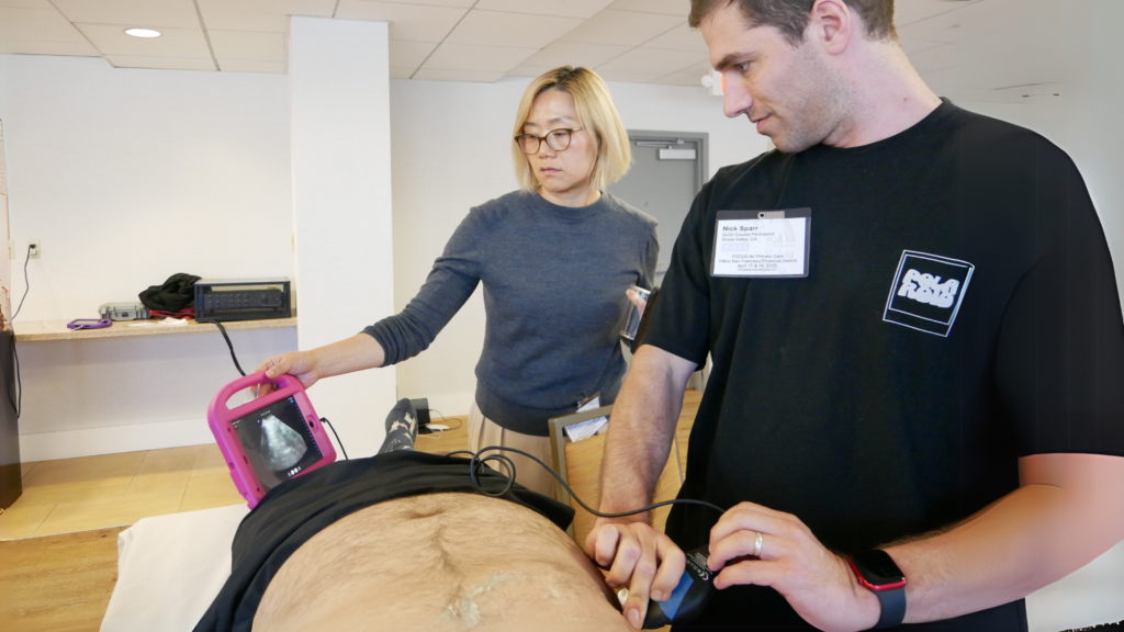
Professional ultrasound organizations publish practice parameters, training guidance, and accreditation standards because image quality and competency directly affect clinical reliability. [9][10]
The Goal Is the Right Test, Not Simply Less Imaging
Radiation avoidance matters, but it is only one part of imaging safety. A delayed diagnosis, missed injury, or incomplete evaluation can be more harmful than the radiation from a properly indicated CT scan. Conversely, ordering CT automatically when a focused bedside ultrasound can provide the needed answer may expose the patient to avoidable radiation, cost, transport, and delay.
The best imaging strategy uses each modality for what it does well:
- Use POCUS to answer focused, time-sensitive questions at the bedside.
- Use CT when comprehensive detail or definitive cross-sectional imaging is required.
- Use both when POCUS guides immediate care and CT completes the diagnostic evaluation.
- Build systems for training, documentation, quality assurance, and expert feedback so POCUS findings can be trusted and limitations are recognized.
Clinicians who understand both the capabilities and boundaries of POCUS are better positioned to choose imaging deliberately, minimize avoidable risk, and escalate without delay when a patient needs more.