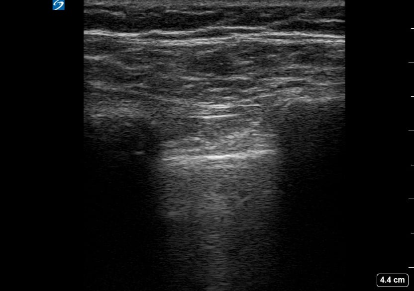
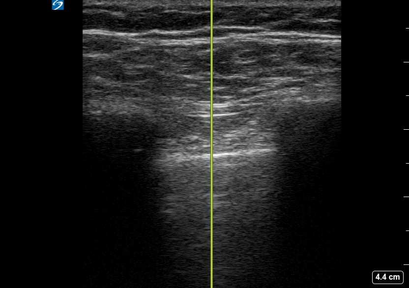
A pneumothorax, or collapsed lung, occurs when air leaks into the space between the lung and chest wall, causing the lung to partially or completely deflate. In a medical setting, prompt diagnosis is crucial to prevent respiratory distress. Ultrasound is a valuable tool for quick assessment, revealing characteristic signs like the absence of lung sliding or the presence of a “barcode” or “stratosphere” sign in M-mode, aiding rapid clinical decisions.
Ultrasound imaging for pneumothorax offers a non-invasive, real-time method for detection, especially in emergency and critical care. Key ultrasound findings guide clinicians in identifying this potentially life-threatening condition, facilitating timely intervention and improved patient outcomes. Understanding these signs is essential for medical professionals utilizing point-of-care ultrasound for lung pathology.