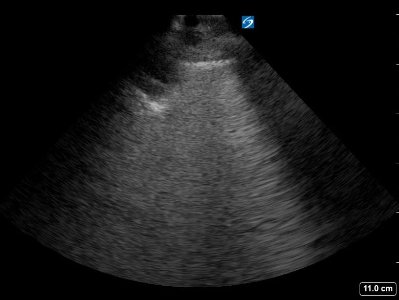
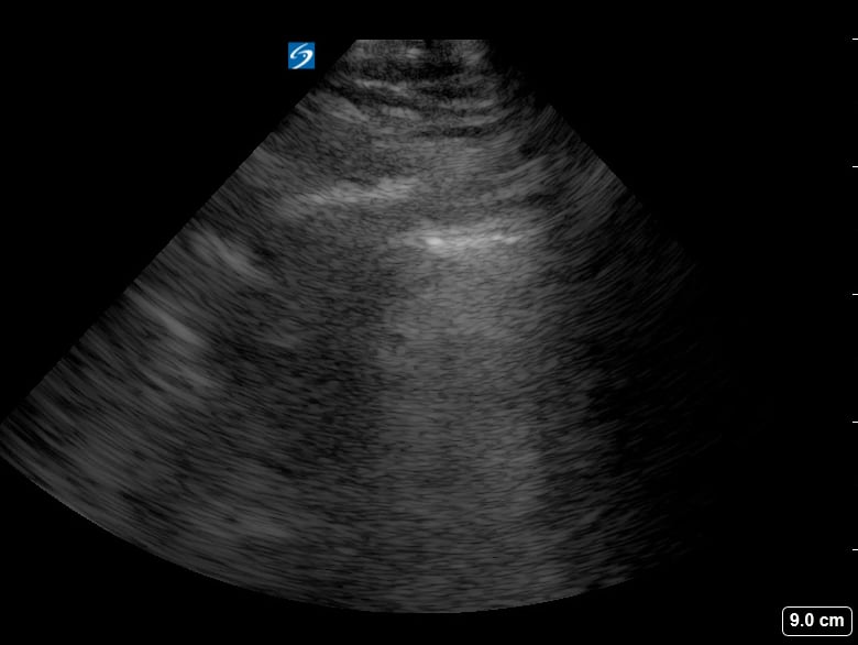
Subpleural consolidation in lung ultrasound refers to an area of lung tissue solidification located directly beneath the pleura, the lining of the lungs. This finding is critical in medical imaging, often indicating inflammatory processes like pneumonia, atelectasis, or pulmonary infarct. Ultrasound excels in visualizing these superficial lung changes due to its high resolution in the near field, making it a valuable tool for bedside assessment and monitoring of lung pathologies.
Identifying subpleural consolidation is crucial for accurate diagnosis and patient management. Its presence can guide clinicians in differentiating various lung conditions, especially in emergency and critical care settings where rapid assessment is vital. Ultrasound’s non-ionizing nature and portability further enhance its utility for repeated evaluations and monitoring of lung disease progression.