Question Bank - Cardiac
Comprehensive Question Bank
Quiz Summary
0 of 126 Questions completed
Questions:
Information
You have already completed the quiz before. Hence you can not start it again.
Quiz is loading...
You must sign in or sign up to start the quiz.
You must first complete the following:
Results
Results
0 of 126 Questions answered correctly
Your time:
Time has elapsed
You have reached 0 of 0 point(s), (0)
Earned Point(s): 0 of 0, (0)
0 Essay(s) Pending (Possible Point(s): 0)
Categories
- Not categorized 0%
- Abdominal Aorta Quiz 0%
- Basics 0%
- 1
- 2
- 3
- 4
- 5
- 6
- 7
- 8
- 9
- 10
- 11
- 12
- 13
- 14
- 15
- 16
- 17
- 18
- 19
- 20
- 21
- 22
- 23
- 24
- 25
- 26
- 27
- 28
- 29
- 30
- 31
- 32
- 33
- 34
- 35
- 36
- 37
- 38
- 39
- 40
- 41
- 42
- 43
- 44
- 45
- 46
- 47
- 48
- 49
- 50
- 51
- 52
- 53
- 54
- 55
- 56
- 57
- 58
- 59
- 60
- 61
- 62
- 63
- 64
- 65
- 66
- 67
- 68
- 69
- 70
- 71
- 72
- 73
- 74
- 75
- 76
- 77
- 78
- 79
- 80
- 81
- 82
- 83
- 84
- 85
- 86
- 87
- 88
- 89
- 90
- 91
- 92
- 93
- 94
- 95
- 96
- 97
- 98
- 99
- 100
- 101
- 102
- 103
- 104
- 105
- 106
- 107
- 108
- 109
- 110
- 111
- 112
- 113
- 114
- 115
- 116
- 117
- 118
- 119
- 120
- 121
- 122
- 123
- 124
- 125
- 126
- Current
- Review / Skip
- Answered
- Correct
- Incorrect
-
Question 1 of 126
1. Question
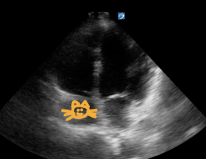
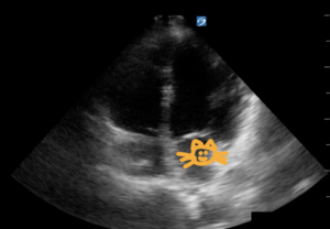
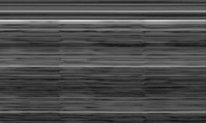
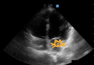
The following image of the heart in the apical 4 chamber window is in which mode?
CorrectIncorrect -
Question 2 of 126
2. Question
Which of the following positioning techniques can help you visualize the abdominal aorta?
CorrectIncorrect -
Question 3 of 126
3. Question
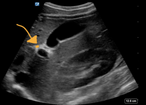
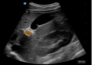
A 45 y/o female is suffering from abdominal pain after meals. What ultrasound mode is used to scan her gallbladder in the image below?
CorrectIncorrect -
Question 4 of 126
4. Question
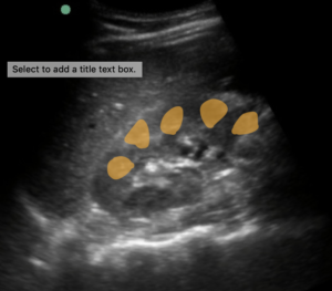
A 45 y/o with a history of kidney stones presents with acute onset of L flank pain. A renal ultrasound shows finding consistent with mild hydronephrosis. To determine whether or not what appears to be hydronephrosis are actually the renal vessels, what machine mode is used below to troubleshoot this potential pitfall?
CorrectIncorrect -
Question 5 of 126
5. Question
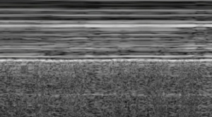
A 23 year old male fell off a bicycle and complains of rib pain, shortness of breath and abdominal pain. A focused assessment of sonography in trauma is performed (FAST exam). Which artifact seen above the diaphragm suggests that the patient does NOT have a hemothorax?
CorrectIncorrect -
Question 6 of 126
6. Question
Where are 90% of AAA’s located?
CorrectIncorrect -
Question 7 of 126
7. Question
Which of the following descriptions of ultrasound frequency and probe selection is most accurate?
CorrectIncorrect -
Question 8 of 126
8. Question
If you are unable to find the gall bladder with a supine patient, what should you do?
CorrectIncorrect -
Question 9 of 126
9. Question
A 23 year old male is involved in an accident falling forward off of his bicycle hitting his abdomen on the handlebars. He is in severe abdominal pain and has visible bruising over his left flank and around his umbilicus. You perform an ultrasound exam which shows free fluid in the left upper quadrant between the diaphragm and the spleen concern for a splenic injury. How do you describe the appearance of fluid on an ultrasound and what happens to sound waves that create this appearance?
CorrectIncorrect -
Question 10 of 126
10. Question
What is the most reliable sonographic landmark to localize the abdominal aorta?
CorrectIncorrect -
Question 11 of 126
11. Question
Sometimes you have to exert some force with the probe to see if the vein will collapse, especially for fluffier or for more musclebound patients. How do you know how hard is hard enough to compress for either a positive or negative POCUS DVT scan?
CorrectIncorrect -
Question 12 of 126
12. Question
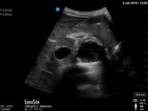
This patient’s abdominal aorta measures 4.3cm at its greatest diameter. Do they have a abdominal aortic aneurysm by definition?
CorrectIncorrect -
Question 13 of 126
13. Question
An iliac aneurysm is by definition greater than how many centimeters wide?
CorrectIncorrect -
Question 14 of 126
14. Question
The following image is taken in the sagittal plane in the abdominal right upper quadrant. In this view, the sonographer is evaluating Morrison's Pouch, the space between the liver and kidney, to evaluate for free fluid concerning for hemoperitoneum in the setting of trauma. In which direction, relative to the patient, is the probe marker pointing?
CorrectIncorrect -
Question 15 of 126
15. Question
From where to where should you correctly measure this abdominal aorta?
CorrectIncorrect -
Question 16 of 126
16. Question
A 75 year old male with long standing history of smoking is seen in your clinic. The curvilinear probe is placed in the transverse plane in the abdominal midline above the umbilicus. The following image suggests:
CorrectIncorrect -
Question 17 of 126
17. Question
To evaluate for a DVT, the linear probe is used in the popliteal region. In the following video, what probe movement is used to rule out the presence of a popliteal DVT?
CorrectIncorrect -
Question 18 of 126
18. Question
A 16 year old male presents with RLQ pain, nausea, vomiting and positive straight leg raise. The ultrasound probe is placed in the RLQ at the point of maximal tenderness for the patient. What sonographic finding is present below?
CorrectIncorrect -
Question 19 of 126
19. Question
A 23 year sexually active male presents to your clinic with testicular pain and swelling. Ultrasound exam below suggests what finding?
CorrectIncorrect -
Question 20 of 126
20. Question
72 y/o male with history of COPD presents with SOB and dyspnea on exertion. A bedside lung ultrasound is performed showing a bright hyperechoic line representing the pleura. It appears to be sliding therefore ruling out the presence of pneumothorax at this intercostal space. Which finding below suggests the lung if full of air at this intercostal space?
CorrectIncorrect -
Question 21 of 126
21. Question
In the sagittal plane, the probe marker points in which direction?
CorrectIncorrect -
Question 22 of 126
22. Question
A 45 yo presents to your clinic with shortness of breath, orthopnea, bilateral leg swelling and recent weight gain. Which artifact present below would you expect to find on this patient's ultrasound?
CorrectIncorrect -
Question 23 of 126
23. Question
The presence of the liver superior to the diaphragm is an example of what type of artifact?
CorrectIncorrect -
Question 24 of 126
24. Question
A 45 y/o female with a history of breast cancer presents to your clinic complaining of shortness of breath and pleuritic chest pain. She has diminished breath sounds in the right lower hemithorax. What finding is present below?
CorrectIncorrect -
Question 25 of 126
25. Question
A 47 y/o male with a history of peptic ulcer disease presents with worsening pain in the right upper quadrant after meals. An H2 blocker and PPI provide him with no relief. His clinician performs the ultrasound below. Applying pressure to the probe causes him more pain. What artifact present below confirms the patient's diagnosis?
CorrectIncorrect -
Question 26 of 126
26. Question
A 65 year old diabetic experiences sudden onset of diminished vision in her left eye. She describes it as "seeing flashes and floaters." A bedside US performed showed the following:
What is the diagnosis?
CorrectIncorrect -
Question 27 of 126
27. Question
You measure the inner wall to inner wall of the abdominal aorta?
CorrectIncorrect -
Question 28 of 126
28. Question
What medications are indicated for this patient with leg swelling? (This scan is being done behind the patient's knee)
CorrectIncorrect -
Question 29 of 126
29. Question
You are considering the diagnosis of an aortic dissection in patient with searing chest pain that radiates to the back. You have just scanned the entire course of the abdominal aorta to the proximal iliac arteries and you do not see a dissection flap. If you want to rule out an aortic dissection, your next step is to:
CorrectIncorrect -
Question 30 of 126
30. Question
What pathology is seen here?
CorrectIncorrect -
Question 31 of 126
31. Question
From inguinal area down to the knee crease, what are the three key junctions/branches of the femoral vein?
CorrectIncorrect -
Question 32 of 126
32. Question
Point of care ultrasound (POCUS) differs from comprehensive radiology ultrasound in that POCUS, we seek to answer focused clinically relevant questions at the bedside. The focused cardiac echo answers which focused questions?
CorrectIncorrect -
Question 33 of 126
33. Question
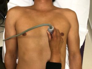
The Apical 4 Chamber view is often the most difficult cardiac view to obtain. Which patient position helps obtain an apical 4 Chamber view?
CorrectIncorrect -
Question 34 of 126
34. Question
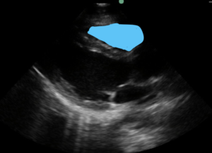
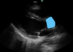
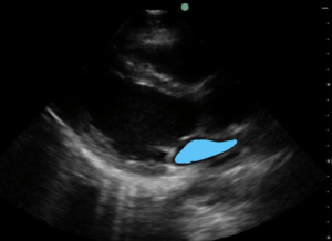
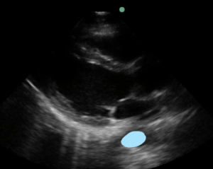
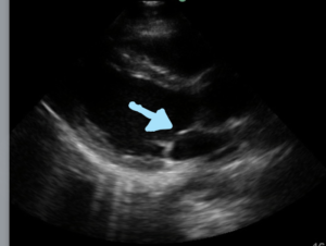
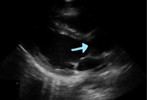
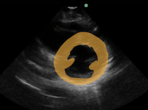
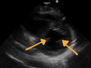
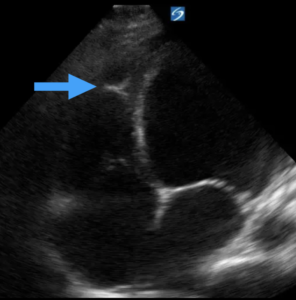
A 45 y/o male with a long standing history of methamphetamine use presents to your clinic with several weeks of shortness of breath. He denies any coughing and fevers. A bedside ultrasound is performed revealing the following findings on parasternal long axis view. What findings are present below?
CorrectIncorrect -
Question 35 of 126
35. Question
In addition to compression, color and pulse wave doppler are necessary to rule in or out the diagnosis of lower extremity DVT.
CorrectIncorrect -
Question 36 of 126
36. Question
If you have having difficulty visualizing the common femoral vessels when scanning the inguinal area, what position change can help?
CorrectIncorrect -
Question 37 of 126
37. Question
As you are scanning down the distal thigh towards the knee, which techniques are most helpful to be able to visualize the femoral vein as it courses deeper toward the bone?
CorrectIncorrect -
Question 38 of 126
38. Question
When you are scanning the popliteal area from behind the knee crease, which vessel is typically on top (closest to the probe)? This scan of the popliteal region suggests what finding?
CorrectIncorrect -
Question 39 of 126
39. Question
What landmark is the most important to see in the inguinal area of the DVT exam? The exam of this area shown below suggests:
CorrectIncorrect -
Question 40 of 126
40. Question
A 27 year old healthy male fell off of a skateboard after attempting to do a jump. He is complaining of abdominal pain and has bruising around the left upper part of his abdomen just beneath the ribs. His BP is 90/60 and HR 118.
Given the findings above of the left upper quadrant, what would be your next step in management?CorrectIncorrect -
Question 41 of 126
41. Question
A 35 year old was roller blading down a hill, tripped and fell and hit her chest. She denies any head trauma, headache, neck pain, abdominal pain or loss of conscious. She hit her chest and feels lots of pain with some shortness of breath. An Extended FAST exam was performed. The finding above suggests:
CorrectIncorrect -
Question 42 of 126
42. Question
What are the most important sonographic questions when ultrasounding the gallbladder?
CorrectIncorrect -
Question 43 of 126
43. Question
A 55 year old female sees you in clinic after several weeks of right upper quadrant pain with meals. She denies fevers, chills, nausea, vomiting. The ultrasound below suggests?
CorrectIncorrect -
Question 44 of 126
44. Question
The Focused Assessment with Sonography in Trauma (FAST) exam has largely replaced diagnostic peritoneal lavage in acute trauma. The FAST exam seeks to answer which two focused questions:
CorrectIncorrect -
Question 45 of 126
45. Question
What is the most reliable sonographic landmark when trying to locate the abdominal aorta?
CorrectIncorrect -
Question 46 of 126
46. Question
What axis and procedural approach is shown below?
CorrectIncorrect -
Question 47 of 126
47. Question
A 35 year wrestler thinks he stepped on something and feels like something is stuck under his foot. The ultrasound below shows a foreign body. What technique can be useful to better visualize foreign bodies near the surface of the skin?
CorrectIncorrect -
Question 48 of 126
48. Question
A 30 year male with acute L flank pain radiating to the groin and hematuria presents to your clinic. Which probe do you select to scan the kidney and why?
CorrectIncorrect -
Question 49 of 126
49. Question
A 35 year old male with acute onset of L flank pain radiating to the groin presents to your office. He has had multiple CT scans in the past which have shown ureteral stones. He has never had urological interventions as the stones have always passed on their own. He denies any fevers & chills. You perform a bedside ultrasound which shows the following image. What finding is present?
CorrectIncorrect -
Question 50 of 126
50. Question
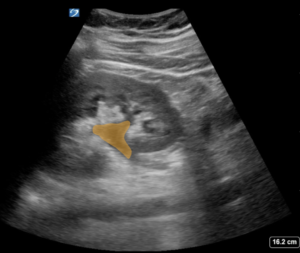
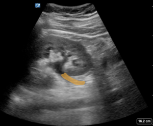
You perform an ultrasound of the right kidney and visualize hypoechoic area in the renal pelvis. You suspect the presence of hydronephrosis.
For confirmation, you use color mode and see the following image.
What do these findings suggest?
CorrectIncorrect -
Question 51 of 126
51. Question
The Rapid Ultrasound In Shock (RUSH) exam is a rapid sonographic approach to narrow the differential for a patient's cause of shock. What are the components of the RUSH exam?
CorrectIncorrect -
Question 52 of 126
52. Question
What best describes the axis of the vessel and the procedural approach.
CorrectIncorrect -
Question 53 of 126
53. Question
A 23 year female presents to the emergency department with abdominal pain and vaginal bleeding. She is unsure of when her last menstrual period and she is sexually active. She denies any history of trauma. She is found to be hypotensive and tachycardic and tender to palpation. Her RUSH exam shows the following findings:
What is the most likely cause of this patient's shock?
CorrectIncorrect -
Question 54 of 126
54. Question
A 65 year old female with a history of hypertension off medications for the last few months presents after progressive shortness of breath and orthopnea. She denies any chest pain, fevers. Her BP if found to be 80/40 and her HR 115. Her RUSH exam shows the following:
What is the most likely cause of the patient's shock?
CorrectIncorrect -
Question 55 of 126
55. Question
What the sensitivity and specificity of POCUS for the diagnosis of DVT?
CorrectIncorrect -
Question 56 of 126
56. Question
A 15 year old male presents with a bump on his arm, pain, redness and swelling. He has been on antibiotics for 5 days and but it is not getting any better. There is no spontaneous drainage. Your ultrasound exam over the area of swelling suggests the presence of:
CorrectIncorrect -
Question 57 of 126
57. Question
The use of ultrasound is useful to distinguish cellulitis from abscess and can minimize the number of dry taps performed.
CorrectIncorrect -
Question 58 of 126
58. Question
The saphenous vein joins with the common femoral vein _____; the popliteal vein is _____ the popliteal artery.
CorrectIncorrect -
Question 59 of 126
59. Question
Does this patient have a DVT?CorrectIncorrect -
Question 60 of 126
60. Question
This 38 year old construction worker patient presents with shoulder pain x 7 days, specifically with difficulty and pain abducting arm. Here is your POCUS scan of his supraspinatus tendon. What is the best next step for this patient?CorrectIncorrect -
Question 61 of 126
61. Question
A 45 year old female with obesity is experiencing pain in the right upper quadrant after meals. You decide to do an ultrasound to look for gallstones. What are the characteristics of the probe that will allow you to best visualize the gallbladder?
CorrectIncorrect -
Question 62 of 126
62. Question
A 17 year female with a history of diabetes presents to you with fever, pain, swelling, warmth and redness in the right leg. Her ultrasound is negative for DVT though shows the following image. What does this finding suggest?
CorrectIncorrect -
Question 63 of 126
63. Question
CorrectIncorrect -
Question 64 of 126
64. Question
CorrectIncorrect -
Question 65 of 126
65. Question
CorrectIncorrect -
Question 66 of 126
66. Question
CorrectIncorrect -
Question 67 of 126
67. Question
CorrectIncorrect -
Question 68 of 126
68. Question
CorrectIncorrect -
Question 69 of 126
69. Question
CorrectIncorrect -
Question 70 of 126
70. Question
CorrectIncorrect -
Question 71 of 126
71. Question
CorrectIncorrect -
Question 72 of 126
72. Question
CorrectIncorrect -
Question 73 of 126
73. Question
CorrectIncorrect -
Question 74 of 126
74. Question
CorrectIncorrect -
Question 75 of 126
75. Question
CorrectIncorrect -
Question 76 of 126
76. Question
CorrectIncorrect -
Question 77 of 126
77. Question
CorrectIncorrect -
Question 78 of 126
78. Question
CorrectIncorrect -
Question 79 of 126
79. Question
CorrectIncorrect -
Question 80 of 126
80. Question
CorrectIncorrect -
Question 81 of 126
81. Question
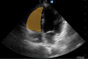
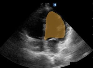
A 45 year old female with a long standing history of hypertension and methamphetamine use presents to clinic for follow up after recent hospitalization for congestive heart failure. Her shortness of breath has improved. Considering the focused cardiac questions, what pathological findings are present in the image below?
CorrectIncorrect -
Question 82 of 126
82. Question
A 63 year old female with a long standing history of hypertension, diabetes, coronary artery disease presents to clinic for follow up after recent hospitalization for congestive heart failure. Her shortness of breath has improved. Considering the focused cardiac questions, what pathological findings are present in the image below?
CorrectIncorrect -
Question 83 of 126
83. Question
A 53 year old female with a history of breast cancer undergoing chemotherapy presents with acute onset of shortness of breath and chest pain. Her bedside echo shows the following finding:
Based on the ultrasound above, what is your most likely diagnosis?
CorrectIncorrect -
Question 84 of 126
84. Question
A 65 year old male with a history of congestive heart failure, COPD, hypertension, and diabetes presents to the clinic with shortness of breath. His vital signs are within normal limits. He reports he gets more short of breath with minimal exertion. He has no chest pain.
His bedside cardiac and lung ultrasound shows the following findings:
Right Thorax:
Left Thorax:
What is your most likely diagnosis for this patient's acute dyspnea?
CorrectIncorrect -
Question 85 of 126
85. Question
A 63 year old female with a history of COPD, hypertension, and diabetes presents to the clinic with cough, subjective fever, and shortness of breath. Her CXR shows a right lower lobe pneumonia.
Her cardiac echo shows what finding?
CorrectIncorrect -
Question 86 of 126
86. Question
CorrectIncorrect -
Question 87 of 126
87. Question
CorrectIncorrect -
Question 88 of 126
88. Question
CorrectIncorrect -
Question 89 of 126
89. Question
CorrectIncorrect -
Question 90 of 126
90. Question
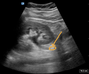
A 23 year old female with acute onset of left flank pain radiating to the groin presents to clinic. She has urinalysis with red blood cells. You perform the ultrasound which shows the image below.
What finding is highlighted in the image below?
CorrectIncorrect -
Question 91 of 126
91. Question
A 23 year old male presents to the emergency department with left flank pain. He has a long history of chronic pain. He is complaining of pain mostly in his back. His urinalysis shows no signs of acute infection or hematuria.
A bedside ultrasound of the left kidney shows the following findings.
What conclusion can you draw of the left kidney?
CorrectIncorrect -
Question 92 of 126
92. Question
A 65 year old female with a history of hypertension and diabetes presents with flank pain, weight loss, and hematuria. Her vital signs are within normal limits. Her WBC, creatinine, platelets and hemoglobin are normal.
Her bedside ultrasound shows the following finding of the let kidney.
What is an appropriate next step in management?
CorrectIncorrect -
Question 93 of 126
93. Question
A 45 year old female presents with acute flank pain radiating to the groin and hematuria and you suspect an acute ureteral obstruction from a kidney stone.
Which of the following are indications for obtaining further imaging such as a CT scan?
CorrectIncorrect -
Question 94 of 126
94. Question
A 35 year old female presents with right flank pain radiating to the groin, nausea, vomiting and hematuria. Your history and physical exam is suspicious for an obstructing kidney stone. Her vitals are within normal limits. She has no tenderness in the RLQ or RUQ. Her gallbladder ultrasound shows no signs of stones or sonographic Murphy's sign. Her WBC count, CRP, Hg, PLT, Renal Function are all normal. Her urinalysis shows no signs of infection.
Her right and left kidney ultrasound show the following:
What is an appropriate next step in management?
CorrectIncorrect -
Question 95 of 126
95. Question
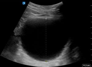
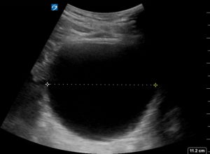
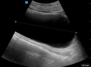
A 63 yo male presents with difficulty urinating, lower abdominal fullness, and pain. His creatinine is elevated from 0.7 to 3.5.
A bedside ultrasound of his bladder following a void trial shows the following:
What is the more likely diagnosis and what is the most appropriate treatment?
CorrectIncorrect -
Question 96 of 126
96. Question
What focused questions are answered by the Focused Assessment with Sonography For Trauma exam?
CorrectIncorrect -
Question 97 of 126
97. Question
What focused questions are answered by the extended FAST exam?
CorrectIncorrect -
Question 98 of 126
98. Question
A 46 year old female is involved in a motor vehicle accident. She was rear ended and hit her chest on the steering wheel. She was wearing a seatbelt. She is complaining of pain over the ribs. Her Xray shows no signs of rib fracture.
What conclusion can be drawn from the following ultrasound?
CorrectIncorrect -
Question 99 of 126
99. Question
A 35 year old female fell off of her bicycle and is complaining of abdominal pain. She has bruising throughout her abdomen and has significant pain in the left upper part of the abdomen. Her FAST exam shows the following:
What conclusion can be drawn?CorrectIncorrect -
Question 100 of 126
100. Question
T/F. In the FAST exam, it is sufficient to evaluate the pelvic view in the transverse plane.
CorrectIncorrect -
Question 101 of 126
101. Question
T/F. In the FAST exam, it is sufficient to evaluate the RUQ view between the liver and kidney (Morrison's pouch).
CorrectIncorrect -
Question 102 of 126
102. Question
A 21 year old male falls off of his roller skates while going down hill. He presents with over his clavicle and is found to have a fracture. An extended FAST exam is performed.
What conclusion can be drawn based on this ultrasound in the right lung apex?
CorrectIncorrect -
Question 103 of 126
103. Question
A 48 year old male is involved in a motorcycle accident and is complaining of rib pain and shortness of breath. A bedside ultrasound is performed of the lungs and shows the following finding in M-Mode.
Based on the above, what conclusion can you draw?
CorrectIncorrect -
Question 104 of 126
104. Question
A 42 year old female fell off her bike on her way to work. She has abrasions on her arms and legs and across her chest. She is complaining of pain in her ribs and shortness of breath.
An ultrasound of her lungs showing the following:
Right Mid-Clavicular Line
Left Mid-Clavicular Line
CorrectIncorrect -
Question 105 of 126
105. Question
A 57 year old male involved in a high speed motor vehicle accident on the highway. He is brought to the emergency department with multiple contusions to the chest and shortness of breath. An extended FAST exam is performed. What conclusion can be drawn based on the following finding:
Right Costophrenic Angle
Left Costophrenic Angle
CorrectIncorrect -
Question 106 of 126
106. Question
True/False. It is adequate to scan the lower extremity venous at two points, the femoral and the popliteal veins, in order to rule out DVT.
CorrectIncorrect -
Question 107 of 126
107. Question
CorrectIncorrect -
Question 108 of 126
108. Question
CorrectIncorrect -
Question 109 of 126
109. Question
CorrectIncorrect -
Question 110 of 126
110. Question
CorrectIncorrect -
Question 111 of 126
111. Question
CorrectIncorrect -
Question 112 of 126
112. Question
CorrectIncorrect -
Question 113 of 126
113. Question
A 35 year old female presents to your clinic with unilateral leg pain and swelling. An ultrasound of the painful leg is performed to evaluate for a deep venous thrombosis. The highlighted image below is non compressible.
What is the appropriate next in management?
CorrectIncorrect -
Question 114 of 126
114. Question
A 48 year old female presents to your clinic complaining of leg pain and swelling. She recently returned 3 days ago after going on vacation to South Africa (a 13 hour plane ride). You perform a bedside ultrasound that shows the following:
Right popliteal fossa
What is an appropriate next stop in management?CorrectIncorrect -
Question 115 of 126
115. Question
A 65 year old male underwent an open reduction external fixation orthopedic surgery after fracturing his right hip 2 weeks ago. He is seen in clinic for a follow up appointment and noticed that his right leg has been more swollen than usual.
A bedside ultrasound in clinic is performed and shows the following finding from the right popliteal fossa:
What is the most appropriate next step in management?
CorrectIncorrect -
Question 116 of 126
116. Question
A 57 year old male with a history of homelessness and chronic alcoholism presents complaining of pain and swelling in his legs, primarily his left leg. He says his legs are always swollen because he has a hard time elevating them. He has no history of shortness of breath, orthopnea, or exertional dyspnea. You perform a bedside ultrasound of the left leg to evaluate him for a deep venous thrombosis.
Based on this ultrasound below the left inguinal canal, what is the next appropriate step in management?
CorrectIncorrect -
Question 117 of 126
117. Question
A 73 year old female presents to your clinic with pain radiating from her right gluteus down her right leg. She also noticed that the leg appears slightly more swollen. On her exam, the legs appear symmetric. The patient also reports she was treated for a deep venous thrombosis over 10 years ago which she developed after having hip surgery.
You perform a bedside ultrasound that shows the following finding:
What is the most appropriate next step in management for this patient?
CorrectIncorrect -
Question 118 of 126
118. Question
CorrectIncorrect -
Question 119 of 126
119. Question
CorrectIncorrect -
Question 120 of 126
120. Question
CorrectIncorrect -
Question 121 of 126
121. Question
CorrectIncorrect -
Question 122 of 126
122. Question
A 52 year old female with a history of obesity and diabetes, presents for the 3rd time for upper abdominal pain. She says it gets worse after she eats. She was treated with omeprazole and ranitidine as well as treated for H. Pylori however her symptoms have not improved. Her vital signs are normal. Her labs show a mild elevation aspartate transaminase (AST) and alanine transaminase (ALT) of 110, 115 respectively and her WBC count is normal.
After performing your physical exam, your bedside ultrasound of the RUQ shows the following. Upon visualizing this finding, gentle pressure applied causes the patient discomfort.
What is the most likely diagnosis?
CorrectIncorrect -
Question 123 of 126
123. Question
A 45 year old female G5P4 with a history of obesity, presents for her 4th visit for upper abdominal pain. She says it gets worse after she eats. She was treated with omeprazole and ranitidine as well as treated for H. Pylori however her symptoms have not improved. Her vital signs are normal.
After performing your physical exam, your bedside ultrasound of the RUQ shows the following. Upon visualizing this finding, gentle pressure applied causes the patient discomfort.
What is the most appropriate next step in patient management?
CorrectIncorrect -
Question 124 of 126
124. Question
CorrectIncorrect -
Question 125 of 126
125. Question
CorrectIncorrect -
Question 126 of 126
126. Question
CorrectIncorrect

