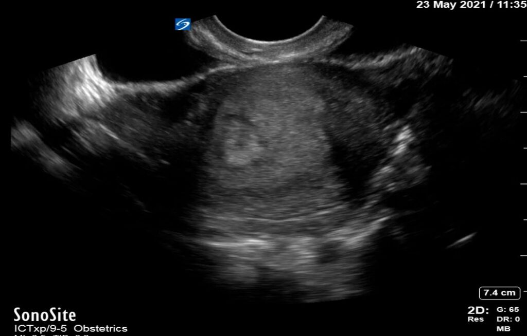
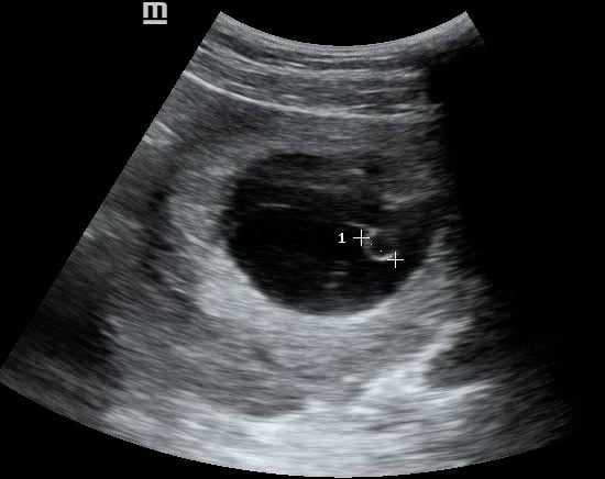
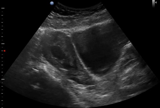
In OBGYN ultrasound, the yolk sac is a crucial early gestational structure, appearing as a small, round, anechoic fluid-filled sac within the chorionic cavity. Typically visible from 5-10 weeks of gestation, its presence confirms an intrauterine pregnancy and is a vital indicator of normal embryonic development. The yolk sac provides early nourishment to the embryo before the placenta fully forms.
Its proper visualization and size are important for dating the pregnancy and assessing viability. Abnormalities in yolk sac appearance, such as irregular shape or absent visualization when expected, can suggest potential complications or early pregnancy failure. Therefore, precise identification and evaluation of the yolk sac are fundamental for accurate diagnostic ultrasound in obstetrics.